 Movie
Movie Controller

[English] 日本語
 Yorodumi
- PDB-2q4g: Ensemble refinement of the protein crystal structure of human rib... -
+ Open data

- Basic information
| Entry | Database: PDB / ID: 2q4g | ||||||
|---|---|---|---|---|---|---|---|
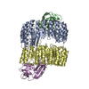
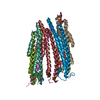
| Title | Ensemble refinement of the protein crystal structure of human ribonuclease inhibitor complexed with ribonuclease I | ||||||
 Components Components |
| ||||||
 Keywords Keywords | HYDROLASE/HYDROLASE INHIBITOR / Ensemble Refinement / Refinement Methodology Development / RIBONUCLEASE-INHIBITOR COMPLEX / LEUCINE-RICH REPEAT / ENZYME-INHIBITOR COMPLEX / Structural Genomics / Protein Structure Initiative / PSI / Center for Eukaryotic Structural Genomics / CESG / HYDROLASE-HYDROLASE INHIBITOR COMPLEX | ||||||
| Function / homology |  Function and homology information Function and homology informationribonuclease inhibitor activity / tRNA stabilization / angiogenin-PRI complex / Developmental Lineage of Pancreatic Acinar Cells / regulation of Arp2/3 complex-mediated actin nucleation / pancreatic ribonuclease / ribonuclease A activity / mRNA catabolic process / regulation of angiogenesis / RNA nuclease activity ...ribonuclease inhibitor activity / tRNA stabilization / angiogenin-PRI complex / Developmental Lineage of Pancreatic Acinar Cells / regulation of Arp2/3 complex-mediated actin nucleation / pancreatic ribonuclease / ribonuclease A activity / mRNA catabolic process / regulation of angiogenesis / RNA nuclease activity / Late endosomal microautophagy / Chaperone Mediated Autophagy / cell migration / lamellipodium / nucleic acid binding / defense response to Gram-positive bacterium / hydrolase activity / extracellular exosome / nucleoplasm / nucleus / plasma membrane / cytosol / cytoplasm Similarity search - Function | ||||||
| Biological species |  Homo sapiens (human) Homo sapiens (human) | ||||||
| Method |  X-RAY DIFFRACTION / Re-refinement using ensemble model / Resolution: 1.954 Å X-RAY DIFFRACTION / Re-refinement using ensemble model / Resolution: 1.954 Å | ||||||
 Authors Authors | Levin, E.J. / Kondrashov, D.A. / Wesenberg, G.E. / Phillips Jr., G.N. / Center for Eukaryotic Structural Genomics (CESG) | ||||||
 Citation Citation | Journal: J.Mol.Biol. / Year: 2007 Title: Inhibition of human pancreatic ribonuclease by the human ribonuclease inhibitor protein. Authors: Johnson, R.J. / McCoy, J.G. / Bingman, C.A. / Phillips Jr., G.N. / Raines, R.T. #1: Journal: Structure / Year: 2007Title: Ensemble refinement of protein crystal structures: validation and application. Authors: Levin, E.J. / Kondrashov, D.A. / Wesenberg, G.E. / Phillips Jr., G.N. | ||||||
| History |
|
- Structure visualization
| Structure viewer | Molecule: MolmilJmol/JSmol |
|---|
- Downloads & links
-Download
| PDBx/mmCIF format | 2q4g.cif.gz | 1.7 MB | Display | PDBx/mmCIF format |
|---|---|---|---|---|
| PDB format | pdb2q4g.ent.gz | 1.5 MB | Display | PDB format |
| PDBx/mmJSON format | 2q4g.json.gz | Tree view | PDBx/mmJSON format | |
| Others |  Other downloads Other downloads |
-Validation report
| Arichive directory | https://data.pdbj.org/pub/pdb/validation_reports/q4/2q4gftp://data.pdbj.org/pub/pdb/validation_reports/q4/2q4g | HTTPS FTP |
|---|
-Related structure data
| Related structure data |  1z7xSC S: Starting model for refinement C: citing same article ( |
|---|---|
| Similar structure data | |
| Other databases |


-Links
 PDBj
PDBj


- Assembly
| Deposited unit | 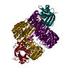
| ||||||||
|---|---|---|---|---|---|---|---|---|---|
| 1 |
| ||||||||
| Unit cell |
| ||||||||
| Number of models | 8 |
-Components
| #1: Protein | Mass: 14728.596 Da / Num. of mol.: 2 Source method: isolated from a genetically manipulated source Source: (gene. exp.) Homo sapiens (human) / Gene: RNASE1, RIB1, RNS1 / Plasmid: pET22b / Species (production host): Escherichia coli / Production host:  #2: Protein | Mass: 50018.355 Da / Num. of mol.: 2 Source method: isolated from a genetically manipulated source Source: (gene. exp.) Homo sapiens (human) / Gene: RNH1, PRI, RNH / Plasmid: pET22b / Species (production host): Escherichia coli / Production host: #3: Chemical | ChemComp-CIT / |   Mass: 192.124 Da / Num. of mol.: 1 / Source method: obtained synthetically / Formula: C6H8O7 Mass: 192.124 Da / Num. of mol.: 1 / Source method: obtained synthetically / Formula: C6H8O7#4: Water | ChemComp-HOH / |  Mass: 18.015 Da / Num. of mol.: 854 / Source method: isolated from a natural source / Formula: H2O Mass: 18.015 Da / Num. of mol.: 854 / Source method: isolated from a natural source / Formula: H2OHas protein modification | Y | |
|---|
-Experimental details
-Experiment
| Experiment | Method: X-RAY DIFFRACTION |
|---|
- Sample preparation
| Crystal | Density Matthews: 2.3 Å3/Da / Density % sol: 46.5 % / Description: AUTHOR USED THE SF DATA FROM ENTRY 1Z7X. |
|---|
-Data collection
| Radiation | Protocol: SINGLE WAVELENGTH / Monochromatic (M) / Laue (L): M / Scattering type: x-ray |
|---|---|
| Radiation wavelength | Relative weight: 1 |
- Processing
| Software |
| ||||||||||||||||||||||||||||||||||||||||||||||||||||||||||||||||||||||
|---|---|---|---|---|---|---|---|---|---|---|---|---|---|---|---|---|---|---|---|---|---|---|---|---|---|---|---|---|---|---|---|---|---|---|---|---|---|---|---|---|---|---|---|---|---|---|---|---|---|---|---|---|---|---|---|---|---|---|---|---|---|---|---|---|---|---|---|---|---|---|---|
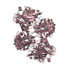
| Refinement | Method to determine structure: Re-refinement using ensemble model Starting model: PDB entry 1Z7X Resolution: 1.954→47.173 Å / Rfactor Rfree error: 0.003 / Data cutoff high absF: 3072992.25 / Data cutoff low absF: 0 / Isotropic thermal model: RESTRAINED / Cross valid method: THROUGHOUT / σ(F): 0 Stereochemistry target values: maximum likelihood using amplitudes Details: This PDB entry is a re-refinement using an ensemble model of the previously deposited single-conformer structure 1z7x and the first data set in the deposited structure factor file for 1z7x ...Details: This PDB entry is a re-refinement using an ensemble model of the previously deposited single-conformer structure 1z7x and the first data set in the deposited structure factor file for 1z7x along with the R-free set defined therein. The coordinates were generated by an automated protocol from an initial model consisting of 8 identical copies of the protein and non-water hetero-atoms assigned fractional occupancies adding up to one, and a single copy of the solvent molecules. Refinement was carried out with all the conformers present simultaneously and with the potential energy terms corresponding to interactions between the different conformers excluded. The helix and sheet records were calculated using coordinates from the first conformer only. The structure visualization program PYMOL is well-suited for directly viewing the ensemble model presented in this PDB file.
| ||||||||||||||||||||||||||||||||||||||||||||||||||||||||||||||||||||||
| Solvent computation | Solvent model: FLAT MODEL / Bsol: 52.277 Å2 / ksol: 0.351 e/Å3 | ||||||||||||||||||||||||||||||||||||||||||||||||||||||||||||||||||||||
| Displacement parameters | Biso mean: 23.8 Å2
| ||||||||||||||||||||||||||||||||||||||||||||||||||||||||||||||||||||||
| Refine analyze |
| ||||||||||||||||||||||||||||||||||||||||||||||||||||||||||||||||||||||
| Refinement step | Cycle: LAST / Resolution: 1.954→47.173 Å
| ||||||||||||||||||||||||||||||||||||||||||||||||||||||||||||||||||||||
| Refine LS restraints |
| ||||||||||||||||||||||||||||||||||||||||||||||||||||||||||||||||||||||
| LS refinement shell | Refine-ID: X-RAY DIFFRACTION / Total num. of bins used: 6
| ||||||||||||||||||||||||||||||||||||||||||||||||||||||||||||||||||||||
| Xplor file |
|
