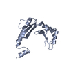
Mass: 8096.414 Da / Num. of mol.: 1 / Source method: obtained synthetically / Details: Chemically synthetized full protein / Source: (synth.) JC polyomavirus / References: UniProt: P03086
-
Experimental details
-
Experiment
Experiment
Method: SOLUTION NMR
NMR experiment
Conditions-ID
Experiment-ID
Solution-ID
Sample state
Spectrometer-ID
Type
1
1
1
anisotropic
1
2D 1H-1H NOESY
1
2
1
anisotropic
1
2D 1H-1H TOCSY
1
3
1
anisotropic
1
2D DQF-COSY
-
Sample preparation
Details
Type: lyophilized powder Contents: 0.2 mM no labelling agnoprotein, trifluoroethanol/water Details: 30% TFE and 200 mM hNaCl ave been added to the solution. The concentration of the sample is 0.2 mM Label: Full agnoprotein / Solvent system: trifluoroethanol/water
Sample
Conc.: 0.2 mM / Component: agnoprotein / Isotopic labeling: no labelling
Sample conditions
Ionic strength: 200 mM NaCl mM / Label: 70/30 (v/v) H2O/TFE / pH: 3 / PH err: 0.2 / Pressure: pa Pa / Temperature: 313 K
-
NMR measurement
NMR spectrometer
Type: Bruker AVANCE III / Manufacturer: Bruker / Model: AVANCE III / Field strength: 600 MHz / Details: cryoprobe
In the structure databanks used in Yorodumi, some data are registered as the other names, "COVID-19 virus" and "2019-nCoV". Here are the details of the virus and the list of structure data.
Jan 31, 2019. EMDB accession codes are about to change! (news from PDBe EMDB page)
EMDB accession codes are about to change! (news from PDBe EMDB page)
The allocation of 4 digits for EMDB accession codes will soon come to an end. Whilst these codes will remain in use, new EMDB accession codes will include an additional digit and will expand incrementally as the available range of codes is exhausted. The current 4-digit format prefixed with “EMD-” (i.e. EMD-XXXX) will advance to a 5-digit format (i.e. EMD-XXXXX), and so on. It is currently estimated that the 4-digit codes will be depleted around Spring 2019, at which point the 5-digit format will come into force.
The EM Navigator/Yorodumi systems omit the EMD- prefix.
Related info.:Q: What is EMD? / ID/Accession-code notation in Yorodumi/EM Navigator
Yorodumi is a browser for structure data from EMDB, PDB, SASBDB, etc.
This page is also the successor to EM Navigator detail page, and also detail information page/front-end page for Omokage search.
The word "yorodu" (or yorozu) is an old Japanese word meaning "ten thousand". "mi" (miru) is to see.
Related info.:EMDB / PDB / SASBDB / Comparison of 3 databanks / Yorodumi Search / Aug 31, 2016. New EM Navigator & Yorodumi / Yorodumi Papers / Jmol/JSmol / Function and homology information / Changes in new EM Navigator and Yorodumi
 Movie
Movie Controller

 Yorodumi
 Open data
 Basic information
 Components
 Keywords
 Function and homology information
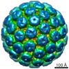 JC polyomavirus
 Authors
 United States, 2items
  Citation
 Structure visualization
 Downloads & links
 Other downloads

 PDBj
PDBj Assembly
 Sample preparation
 Processing