 Movie
Movie Controller

[English] 日本語
 Yorodumi
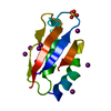
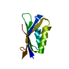
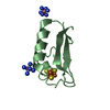
- PDB-5nfm: Crystal structure of YrbA from Sinorhizobium meliloti in complex ... -
+ Open data

- Basic information
| Entry | Database: PDB / ID: 5nfm | ||||||
|---|---|---|---|---|---|---|---|
| Title | Crystal structure of YrbA from Sinorhizobium meliloti in complex with copper. | ||||||
 Components Components | YrbA | ||||||
 Keywords Keywords | LIGASE / BolA / YrbA / histidyl ligation | ||||||
| Function / homology |  Function and homology information Function and homology information | ||||||
| Biological species |  Sinorhizobium meliloti 2011 (bacteria) Sinorhizobium meliloti 2011 (bacteria) | ||||||
| Method |  X-RAY DIFFRACTION / SYNCHROTRON / MOLECULAR REPLACEMENT / Resolution: 0.8 Å X-RAY DIFFRACTION / SYNCHROTRON / MOLECULAR REPLACEMENT / Resolution: 0.8 Å | ||||||
 Authors Authors | Roret, T. / Didierjean, C. | ||||||
 Citation Citation | Journal: Biosci.Rep. / Year: 2020 Title: Sinorhizobium meliloti YrbA binds divalent metal cations using two conserved histidines. Authors: Roret, T. / Alloing, G. / Girardet, J.M. / Perrot, T. / Dhalleine, T. / Couturier, J. / Frendo, P. / Didierjean, C. / Rouhier, N. | ||||||
| History |
|
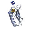
- Structure visualization
| Structure viewer | Molecule: MolmilJmol/JSmol |
|---|
- Downloads & links
-Download
| PDBx/mmCIF format | 5nfm.cif.gz | 62.9 KB | Display | PDBx/mmCIF format |
|---|---|---|---|---|
| PDB format | pdb5nfm.ent.gz | 45.7 KB | Display | PDB format |
| PDBx/mmJSON format | 5nfm.json.gz | Tree view | PDBx/mmJSON format | |
| Others |  Other downloads Other downloads |
-Validation report
| Summary document | 5nfm_validation.pdf.gz | 405.2 KB | Display | wwPDB validaton report |
|---|---|---|---|---|
| Full document | 5nfm_full_validation.pdf.gz | 405.2 KB | Display | |
| Data in XML | 5nfm_validation.xml.gz | 6.8 KB | Display | |
| Data in CIF | 5nfm_validation.cif.gz | 9.3 KB | Display | |
| Arichive directory | https://data.pdbj.org/pub/pdb/validation_reports/nf/5nfmftp://data.pdbj.org/pub/pdb/validation_reports/nf/5nfm | HTTPS FTP |
-Related structure data
| Related structure data | 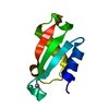 5nfkSC  5nflC S: Starting model for refinement C: citing same article ( |
|---|---|
| Similar structure data |


-Links
 PDBj
PDBj
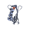
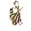
- Assembly
| Deposited unit | 
| ||||||||
|---|---|---|---|---|---|---|---|---|---|
| 1 |
| ||||||||
| Unit cell |
|
-Components
| #1: Protein | Mass: 7955.053 Da / Num. of mol.: 1 Source method: isolated from a genetically manipulated source Source: (gene. exp.) Sinorhizobium meliloti 2011 (bacteria) / Gene: SM2011_c00487 / Production host: |
|---|---|
| #2: Chemical | ChemComp-LI /   Mass: 6.941 Da / Num. of mol.: 1 / Source method: obtained synthetically / Formula: Li Mass: 6.941 Da / Num. of mol.: 1 / Source method: obtained synthetically / Formula: Li |
| #3: Chemical | ChemComp-CU /   Mass: 63.546 Da / Num. of mol.: 1 / Source method: obtained synthetically / Formula: Cu Mass: 63.546 Da / Num. of mol.: 1 / Source method: obtained synthetically / Formula: Cu |
| #4: Water | ChemComp-HOH /  Mass: 18.015 Da / Num. of mol.: 150 / Source method: isolated from a natural source / Formula: H2O Mass: 18.015 Da / Num. of mol.: 150 / Source method: isolated from a natural source / Formula: H2O |
-Experimental details
-Experiment
| Experiment | Method: X-RAY DIFFRACTION / Number of used crystals: 1 |
|---|
- Sample preparation
| Crystal | Density Matthews: 1.94 Å3/Da / Density % sol: 36.75 % |
|---|---|
| Crystal grow | Temperature: 277 K / Method: microbatch Details: 0.7 M lithium sulfate 0.02 M copper acetate 0.1 M Tris-HCl, pH 8.5 |
-Data collection
| Diffraction | Mean temperature: 100 K |
|---|---|
| Diffraction source | Source: SYNCHROTRON / Site: ESRF  / Beamline: BM30A / Wavelength: 0.722307 Å / Beamline: BM30A / Wavelength: 0.722307 Å |
| Detector | Type: ADSC QUANTUM 315r / Detector: CCD / Date: Nov 15, 2014 |
| Radiation | Protocol: SINGLE WAVELENGTH / Monochromatic (M) / Laue (L): M / Scattering type: x-ray |
| Radiation wavelength | Wavelength: 0.722307 Å / Relative weight: 1 |
| Reflection | Resolution: 0.8→32.16 Å / Num. all: 465091 / Num. obs: 66196 / % possible obs: 99.9 % / Redundancy: 7 % / CC1/2: 0.999 / Rmerge(I) obs: 0.084 / Rpim(I) all: 0.034 / Rrim(I) all: 0.091 / Net I/σ(I): 15 |
| Reflection shell | Resolution: 0.8→0.81 Å / Redundancy: 6.8 % / Rmerge(I) obs: 0.856 / Mean I/σ(I) obs: 2.4 / Num. measured obs: 21702 / Num. unique all: 3201 / CC1/2: 0.727 / Rpim(I) all: 0.352 / Rrim(I) all: 0.927 / % possible all: 99 |
- Processing
| Software |
| |||||||||||||||||||||||||||||||||||||||||||||||||||||||||||||||||||||||||||||||||||||||||||||||||||||||||||||||||||||||||||||||||||||||||||||||||||||||||||||||||||||||||||||||
|---|---|---|---|---|---|---|---|---|---|---|---|---|---|---|---|---|---|---|---|---|---|---|---|---|---|---|---|---|---|---|---|---|---|---|---|---|---|---|---|---|---|---|---|---|---|---|---|---|---|---|---|---|---|---|---|---|---|---|---|---|---|---|---|---|---|---|---|---|---|---|---|---|---|---|---|---|---|---|---|---|---|---|---|---|---|---|---|---|---|---|---|---|---|---|---|---|---|---|---|---|---|---|---|---|---|---|---|---|---|---|---|---|---|---|---|---|---|---|---|---|---|---|---|---|---|---|---|---|---|---|---|---|---|---|---|---|---|---|---|---|---|---|---|---|---|---|---|---|---|---|---|---|---|---|---|---|---|---|---|---|---|---|---|---|---|---|---|---|---|---|---|---|---|---|---|---|
| Refinement | Method to determine structure: MOLECULAR REPLACEMENT Starting model: 5NFK Resolution: 0.8→22.425 Å / SU ML: 0.05 / Cross valid method: FREE R-VALUE / σ(F): 1.35 / Phase error: 11.44
| |||||||||||||||||||||||||||||||||||||||||||||||||||||||||||||||||||||||||||||||||||||||||||||||||||||||||||||||||||||||||||||||||||||||||||||||||||||||||||||||||||||||||||||||
| Solvent computation | Shrinkage radii: 0.9 Å / VDW probe radii: 1.11 Å | |||||||||||||||||||||||||||||||||||||||||||||||||||||||||||||||||||||||||||||||||||||||||||||||||||||||||||||||||||||||||||||||||||||||||||||||||||||||||||||||||||||||||||||||
| Refinement step | Cycle: LAST / Resolution: 0.8→22.425 Å
| |||||||||||||||||||||||||||||||||||||||||||||||||||||||||||||||||||||||||||||||||||||||||||||||||||||||||||||||||||||||||||||||||||||||||||||||||||||||||||||||||||||||||||||||
| Refine LS restraints |
| |||||||||||||||||||||||||||||||||||||||||||||||||||||||||||||||||||||||||||||||||||||||||||||||||||||||||||||||||||||||||||||||||||||||||||||||||||||||||||||||||||||||||||||||
| LS refinement shell |
|
