 Movie
Movie Controller

[English] 日本語
 Yorodumi
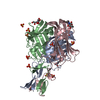
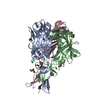
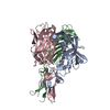
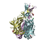
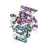
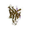
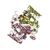
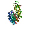
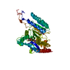
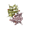
- PDB-5nbh: Structure of the distal domain of mouse adenovirus 2 fibre, P2121... -
+ Open data

- Basic information
| Entry | Database: PDB / ID: 5nbh | ||||||
|---|---|---|---|---|---|---|---|
| Title | Structure of the distal domain of mouse adenovirus 2 fibre, P212121 native | ||||||
 Components Components | Fiber | ||||||
 Keywords Keywords | VIRAL PROTEIN / Triple beta-spiral / 3-bladed propeller / ABCJ-GHID topology / N-acetyl-glucosamine binding | ||||||
| Function / homology |  Function and homology information Function and homology informationadhesion receptor-mediated virion attachment to host cell / viral capsid / cell adhesion / symbiont entry into host cell / host cell nucleus Similarity search - Function | ||||||
| Biological species |  Murine adenovirus 2 Murine adenovirus 2 | ||||||
| Method |  X-RAY DIFFRACTION / SYNCHROTRON / MOLECULAR REPLACEMENT / Resolution: 1.7 Å X-RAY DIFFRACTION / SYNCHROTRON / MOLECULAR REPLACEMENT / Resolution: 1.7 Å | ||||||
 Authors Authors | Singh, A.K. / van Raaij, M.J. | ||||||
 Citation Citation | Journal: J. Gen. Virol. / Year: 2018 Title: Structure and N-acetylglucosamine binding of the distal domain of mouse adenovirus 2 fibre. Authors: Singh, A.K. / Nguyen, T.H. / Vidovszky, M.Z. / Harrach, B. / Benko, M. / Kirwan, A. / Joshi, L. / Kilcoyne, M. / Berbis, M.A. / Canada, F.J. / Jimenez-Barbero, J. / Menendez, M. / Wilson, S. ...Authors: Singh, A.K. / Nguyen, T.H. / Vidovszky, M.Z. / Harrach, B. / Benko, M. / Kirwan, A. / Joshi, L. / Kilcoyne, M. / Berbis, M.A. / Canada, F.J. / Jimenez-Barbero, J. / Menendez, M. / Wilson, S.S. / Bromme, B.A. / Smith, J.G. / van Raaij, M.J. | ||||||
| History |
|
- Structure visualization
| Structure viewer | Molecule: MolmilJmol/JSmol |
|---|
- Downloads & links
-Download
| PDBx/mmCIF format | 5nbh.cif.gz | 144.9 KB | Display | PDBx/mmCIF format |
|---|---|---|---|---|
| PDB format | pdb5nbh.ent.gz | 110.7 KB | Display | PDB format |
| PDBx/mmJSON format | 5nbh.json.gz | Tree view | PDBx/mmJSON format | |
| Others |  Other downloads Other downloads |
-Validation report
| Arichive directory | https://data.pdbj.org/pub/pdb/validation_reports/nb/5nbhftp://data.pdbj.org/pub/pdb/validation_reports/nb/5nbh | HTTPS FTP |
|---|
-Related structure data
| Related structure data |  5n83C  5n8dSC  5nc1C C: citing same article ( S: Starting model for refinement |
|---|---|
| Similar structure data |
-Links
 PDBj
PDBj
- Assembly
| Deposited unit | 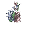
| ||||||||
|---|---|---|---|---|---|---|---|---|---|
| 1 |
| ||||||||
| Unit cell |
|
-Components
| #1: Protein | Mass: 25810.922 Da / Num. of mol.: 3 Source method: isolated from a genetically manipulated source Source: (gene. exp.) Murine adenovirus 2 / Plasmid: pET28a / Production host:  #2: Chemical | ChemComp-GOL / |   Mass: 92.094 Da / Num. of mol.: 1 / Source method: obtained synthetically / Formula: C3H8O3 Mass: 92.094 Da / Num. of mol.: 1 / Source method: obtained synthetically / Formula: C3H8O3#3: Water | ChemComp-HOH / |  Mass: 18.015 Da / Num. of mol.: 672 / Source method: isolated from a natural source / Formula: H2O Mass: 18.015 Da / Num. of mol.: 672 / Source method: isolated from a natural source / Formula: H2O |
|---|
-Experimental details
-Experiment
| Experiment | Method: X-RAY DIFFRACTION / Number of used crystals: 1 |
|---|
- Sample preparation
| Crystal | Density Matthews: 2.12 Å3/Da / Density % sol: 41.95 % |
|---|---|
| Crystal grow | Temperature: 294 K / Method: vapor diffusion, sitting drop / pH: 7.5 Details: 10 mM Tris-HCl, 10 % (w/v) PEG 4000, 0.1 M HEPES-NaOH, 0.1 M magnesium chloride |
-Data collection
| Diffraction | Mean temperature: 100 K |
|---|---|
| Diffraction source | Source: SYNCHROTRON / Site: ESRF  / Beamline: ID29 / Wavelength: 0.9763 Å / Beamline: ID29 / Wavelength: 0.9763 Å |
| Detector | Type: ADSC QUANTUM 315r / Detector: CCD / Date: Feb 21, 2014 |
| Radiation | Protocol: SINGLE WAVELENGTH / Monochromatic (M) / Laue (L): M / Scattering type: x-ray |
| Radiation wavelength | Wavelength: 0.9763 Å / Relative weight: 1 |
| Reflection | Resolution: 1.7→34.1 Å / Num. obs: 68912 / % possible obs: 94.3 % / Redundancy: 3.5 % / Biso Wilson estimate: 14.5 Å2 / CC1/2: 0.983 / Rmerge(I) obs: 0.116 / Net I/σ(I): 6.3 |
| Reflection shell | Resolution: 1.7→1.74 Å / Redundancy: 1.9 % / Rmerge(I) obs: 0.233 / Mean I/σ(I) obs: 2.1 / Num. unique obs: 3722 / CC1/2: 0.857 / % possible all: 69.6 |
- Processing
| Software |
| ||||||||||||||||||||||||||||||||||||||||||||||||||||||||||||||||||||||||||||||||||||||||||||||||||||||||||||||||||||||||||||||||||||||||||||||||||||||||||||||||||||||||||||||||||||||
|---|---|---|---|---|---|---|---|---|---|---|---|---|---|---|---|---|---|---|---|---|---|---|---|---|---|---|---|---|---|---|---|---|---|---|---|---|---|---|---|---|---|---|---|---|---|---|---|---|---|---|---|---|---|---|---|---|---|---|---|---|---|---|---|---|---|---|---|---|---|---|---|---|---|---|---|---|---|---|---|---|---|---|---|---|---|---|---|---|---|---|---|---|---|---|---|---|---|---|---|---|---|---|---|---|---|---|---|---|---|---|---|---|---|---|---|---|---|---|---|---|---|---|---|---|---|---|---|---|---|---|---|---|---|---|---|---|---|---|---|---|---|---|---|---|---|---|---|---|---|---|---|---|---|---|---|---|---|---|---|---|---|---|---|---|---|---|---|---|---|---|---|---|---|---|---|---|---|---|---|---|---|---|---|
| Refinement | Method to determine structure: MOLECULAR REPLACEMENT Starting model: 5N8D Resolution: 1.7→34.05 Å / Cor.coef. Fo:Fc: 0.96 / Cor.coef. Fo:Fc free: 0.93 / SU B: 2.497 / SU ML: 0.08 / Cross valid method: THROUGHOUT / ESU R: 0.105 / ESU R Free: 0.109 / Details: HYDROGENS HAVE BEEN ADDED IN THE RIDING POSITIONS
| ||||||||||||||||||||||||||||||||||||||||||||||||||||||||||||||||||||||||||||||||||||||||||||||||||||||||||||||||||||||||||||||||||||||||||||||||||||||||||||||||||||||||||||||||||||||
| Solvent computation | Ion probe radii: 0.8 Å / Shrinkage radii: 0.8 Å / VDW probe radii: 1.2 Å | ||||||||||||||||||||||||||||||||||||||||||||||||||||||||||||||||||||||||||||||||||||||||||||||||||||||||||||||||||||||||||||||||||||||||||||||||||||||||||||||||||||||||||||||||||||||
| Displacement parameters | Biso mean: 19.75 Å2
| ||||||||||||||||||||||||||||||||||||||||||||||||||||||||||||||||||||||||||||||||||||||||||||||||||||||||||||||||||||||||||||||||||||||||||||||||||||||||||||||||||||||||||||||||||||||
| Refinement step | Cycle: 1 / Resolution: 1.7→34.05 Å
| ||||||||||||||||||||||||||||||||||||||||||||||||||||||||||||||||||||||||||||||||||||||||||||||||||||||||||||||||||||||||||||||||||||||||||||||||||||||||||||||||||||||||||||||||||||||
| Refine LS restraints |
|