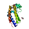
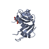
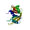
Entry Database : PDB / ID : 5na9Title The X-ray structure of bovine pancreatic ribonuclease incubated in the presence of an excess of carboplatin (1:10 ratio) Ribonuclease pancreatic Keywords / / / Function / homology Function Domain/homology Component
/ / / / / / / / / / / / / / / / Biological species Bos taurus (domestic cattle)Method / / Resolution : 2.07 Å Authors Ferraro, G. / Merlino, A. Journal : J. Inorg. Biochem. / Year : 2017Title : A comparison study on RNase A oligomerization induced by cisplatin, carboplatin and oxaliplatin.Authors : Picone, D. / Donnarumma, F. / Ferraro, G. / Gotte, G. / Fagagnini, A. / Butera, G. / Donadelli, M. / Merlino, A. History Deposition Feb 27, 2017 Deposition site / Processing site Revision 1.0 Aug 16, 2017 Provider / Type Revision 1.1 Jan 17, 2024 Group Advisory / Data collection ... Advisory / Data collection / Database references / Derived calculations / Refinement description Category chem_comp_atom / chem_comp_bond ... chem_comp_atom / chem_comp_bond / database_2 / pdbx_initial_refinement_model / pdbx_struct_conn_angle / pdbx_unobs_or_zero_occ_atoms / struct_conn Item _database_2.pdbx_DOI / _database_2.pdbx_database_accession ... _database_2.pdbx_DOI / _database_2.pdbx_database_accession / _pdbx_struct_conn_angle.ptnr1_auth_comp_id / _pdbx_struct_conn_angle.ptnr1_auth_seq_id / _pdbx_struct_conn_angle.ptnr1_label_asym_id / _pdbx_struct_conn_angle.ptnr1_label_comp_id / _pdbx_struct_conn_angle.ptnr1_label_seq_id / _pdbx_struct_conn_angle.ptnr1_symmetry / _pdbx_struct_conn_angle.ptnr3_auth_comp_id / _pdbx_struct_conn_angle.ptnr3_auth_seq_id / _pdbx_struct_conn_angle.ptnr3_label_asym_id / _pdbx_struct_conn_angle.ptnr3_label_comp_id / _pdbx_struct_conn_angle.ptnr3_label_seq_id / _pdbx_struct_conn_angle.ptnr3_symmetry / _pdbx_struct_conn_angle.value / _struct_conn.pdbx_dist_value / _struct_conn.pdbx_ptnr1_label_alt_id / _struct_conn.pdbx_ptnr2_label_alt_id / _struct_conn.ptnr1_auth_comp_id / _struct_conn.ptnr1_auth_seq_id / _struct_conn.ptnr1_label_asym_id / _struct_conn.ptnr1_label_atom_id / _struct_conn.ptnr1_label_comp_id / _struct_conn.ptnr1_label_seq_id / _struct_conn.ptnr2_auth_comp_id / _struct_conn.ptnr2_auth_seq_id / _struct_conn.ptnr2_label_asym_id / _struct_conn.ptnr2_label_atom_id / _struct_conn.ptnr2_label_comp_id / _struct_conn.ptnr2_symmetry Revision 1.2 Oct 16, 2024 Group / Category / pdbx_modification_feature
Show all Show less
 Movie
Movie Controller

 Yorodumi
 Open data
 Basic information
 Components
 Keywords
 Function and homology information
 X-RAY DIFFRACTION /
  Authors
 Citation
 Structure visualization
 Downloads & links
 Other downloads
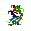


 PDBj
PDBj

 Assembly
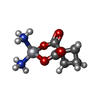
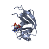
 Mass: 371.248 Da / Num. of mol.: 1 / Source method: obtained synthetically / Formula: C6H12N2O4Pt / Comment: medication, chemotherapy*YM
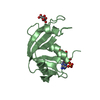
 Mass: 132.905 Da / Num. of mol.: 4 / Source method: obtained synthetically / Formula: Cs
 Mass: 35.453 Da / Num. of mol.: 9 / Source method: obtained synthetically / Formula: Cl
 Mass: 18.015 Da / Num. of mol.: 215 / Source method: isolated from a natural source / Formula: H2O
 Sample preparation
 Processing