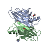
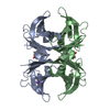
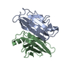
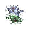
[English] 日本語
Yorodumi
- PDB-5n7c: Human TTR altered conformation from soaking in CuCl2. -

+
Open data


ID or keywords:

Loading...

-
Basic information

Entry
Database: PDB / ID: 5n7c
TitleHuman TTR altered conformation from soaking in CuCl2.
ComponentsTransthyretin
KeywordsTRANSPORT PROTEIN / Copper TTR complex / metal binding / Alzheimer beta-amyloid scavenging
Function / homology
Function and homology information


Retinoid cycle disease events / thyroid hormone binding / The canonical retinoid cycle in rods (twilight vision) / Non-integrin membrane-ECM interactions / purine nucleobase metabolic process / Retinoid metabolism and transport / hormone activity / azurophil granule lumen / Amyloid fiber formation / Neutrophil degranulation ...Retinoid cycle disease events / thyroid hormone binding / The canonical retinoid cycle in rods (twilight vision) / Non-integrin membrane-ECM interactions / purine nucleobase metabolic process / Retinoid metabolism and transport / hormone activity / azurophil granule lumen / Amyloid fiber formation / Neutrophil degranulation / extracellular space / extracellular exosome / extracellular region / identical protein binding
Similarity search - Function
Transthyretin, conserved site / Transthyretin signature 2. / Transthyretin, thyroxine binding site / Transthyretin signature 1. / Transthyretin / Transthyretin/hydroxyisourate hydrolase / Transthyretin/hydroxyisourate hydrolase domain / Transthyretin/hydroxyisourate hydrolase domain superfamily / HIUase/Transthyretin family
Similarity search - Domain/homology
ACETATE ION / COPPER (II) ION / DI(HYDROXYETHYL)ETHER / Transthyretin
Similarity search - Component
Biological speciesHomo sapiens (human)
MethodX-RAY DIFFRACTION / SYNCHROTRON / MOLECULAR REPLACEMENT / Resolution: 2.45 Å
AuthorsCiccone, L. / Savko, M. / Shepard, W. / Stura, E.A.
CitationJournal: Sci Rep / Year: 2018
Title: Copper mediated amyloid-beta binding to Transthyretin.
Authors: Ciccone, L. / Fruchart-Gaillard, C. / Mourier, G. / Savko, M. / Nencetti, S. / Orlandini, E. / Servent, D. / Stura, E.A. / Shepard, W.
History
DepositionFeb 20, 2017Deposition site: PDBE / Processing site: PDBE
Revision 1.0Mar 14, 2018Provider: repository / Type: Initial release
Revision 2.0May 23, 2018Group: Advisory / Data collection ...Advisory / Data collection / Database references / Polymer sequence / Source and taxonomy / Structure summary
Category: entity / entity_poly ...entity / entity_poly / entity_poly_seq / entity_src_nat / pdbx_poly_seq_scheme / pdbx_unobs_or_zero_occ_residues / struct_ref / struct_ref_seq
Item: _entity.formula_weight / _entity_poly.pdbx_seq_one_letter_code ..._entity.formula_weight / _entity_poly.pdbx_seq_one_letter_code / _entity_poly.pdbx_seq_one_letter_code_can / _entity_src_nat.pdbx_end_seq_num / _struct_ref.pdbx_seq_one_letter_code / _struct_ref_seq.db_align_end / _struct_ref_seq.pdbx_auth_seq_align_end / _struct_ref_seq.seq_align_end
Revision 2.1Sep 26, 2018Group: Data collection / Database references / Category: citation / citation_author
Item: _citation.country / _citation.journal_abbrev ..._citation.country / _citation.journal_abbrev / _citation.journal_id_CSD / _citation.journal_id_ISSN / _citation.journal_volume / _citation.page_first / _citation.page_last / _citation.pdbx_database_id_DOI / _citation.pdbx_database_id_PubMed / _citation.title / _citation.year
Revision 2.2Jan 17, 2024Group: Advisory / Data collection ...Advisory / Data collection / Database references / Derived calculations / Refinement description
Category: chem_comp_atom / chem_comp_bond ...chem_comp_atom / chem_comp_bond / database_2 / pdbx_initial_refinement_model / pdbx_struct_conn_angle / pdbx_unobs_or_zero_occ_atoms / struct_conn
Item: _database_2.pdbx_DOI / _database_2.pdbx_database_accession ..._database_2.pdbx_DOI / _database_2.pdbx_database_accession / _pdbx_struct_conn_angle.ptnr1_auth_comp_id / _pdbx_struct_conn_angle.ptnr1_auth_seq_id / _pdbx_struct_conn_angle.ptnr1_label_asym_id / _pdbx_struct_conn_angle.ptnr1_label_atom_id / _pdbx_struct_conn_angle.ptnr1_label_comp_id / _pdbx_struct_conn_angle.ptnr1_label_seq_id / _pdbx_struct_conn_angle.ptnr2_label_alt_id / _pdbx_struct_conn_angle.ptnr2_symmetry / _pdbx_struct_conn_angle.ptnr3_auth_comp_id / _pdbx_struct_conn_angle.ptnr3_auth_seq_id / _pdbx_struct_conn_angle.ptnr3_label_asym_id / _pdbx_struct_conn_angle.ptnr3_label_atom_id / _pdbx_struct_conn_angle.ptnr3_label_comp_id / _pdbx_struct_conn_angle.ptnr3_label_seq_id / _pdbx_struct_conn_angle.value / _struct_conn.pdbx_dist_value / _struct_conn.pdbx_ptnr2_label_alt_id / _struct_conn.ptnr1_auth_asym_id / _struct_conn.ptnr1_auth_comp_id / _struct_conn.ptnr1_auth_seq_id / _struct_conn.ptnr1_label_asym_id / _struct_conn.ptnr1_label_atom_id / _struct_conn.ptnr1_label_comp_id / _struct_conn.ptnr1_label_seq_id / _struct_conn.ptnr2_auth_asym_id / _struct_conn.ptnr2_label_asym_id / _struct_conn.ptnr2_symmetry

-
Structure visualization

Structure viewerMolecule:
MolmilJmol/JSmol

Downloads & links

-
Assembly

Deposited unit
A: Transthyretin
B: Transthyretin
hetero molecules


Theoretical massNumber of molelcules
Total (without water)26,3418
Polymers25,9232
Non-polymers4186
Water3,153175
1
A: Transthyretin
B: Transthyretin
hetero molecules

A: Transthyretin
B: Transthyretin
hetero molecules


Theoretical massNumber of molelcules
Total (without water)52,68216
Polymers51,8464
Non-polymers83612
Water724
TypeNameSymmetry operationNumber
identity operation1_555x,y,z1
crystal symmetry operation2_765-x+2,-y+1,z1
Buried area7150 Å2
ΔGint-67 kcal/mol
Surface area20620 Å2
MethodPISA
Unit cell
Length a, b, c (Å)42.990, 82.540, 67.770
Angle α, β, γ (deg.)90.00, 90.00, 90.00
Int Tables number18
Space group name H-MP21212

-
Components

-
Protein , 1 types, 2 molecules AB

#1: Protein Transthyretin / / ATTR / Prealbumin / TBPA


Mass: 12961.511 Da / Num. of mol.: 2 / Source method: isolated from a natural source / Source: (natural) Homo sapiens (human) / References: UniProt: P02766

-
Non-polymers , 5 types, 181 molecules

#2: Chemical ChemComp-CU / COPPER (II) ION / Copper


Mass: 63.546 Da / Num. of mol.: 3 / Source method: obtained synthetically / Formula: Cu
#3: Chemical ChemComp-PEG / DI(HYDROXYETHYL)ETHER / Diethylene glycol


Mass: 106.120 Da / Num. of mol.: 1 / Source method: obtained synthetically / Formula: C4H10O3
#4: Chemical ChemComp-ACT / ACETATE ION / Acetate


Mass: 59.044 Da / Num. of mol.: 1 / Source method: obtained synthetically / Formula: C2H3O2
#5: Chemical ChemComp-EDO / 1,2-ETHANEDIOL / ETHYLENE GLYCOL / Ethylene glycol


Mass: 62.068 Da / Num. of mol.: 1 / Source method: obtained synthetically / Formula: C2H6O2
#6: Water ChemComp-HOH / water / Water


Mass: 18.015 Da / Num. of mol.: 175 / Source method: isolated from a natural source / Formula: H2O

-
Experimental details

-
Experiment

ExperimentMethod: X-RAY DIFFRACTION / Number of used crystals: 1

-
Sample preparation

CrystalDensity Matthews: 2.37 Å3/Da / Density % sol: 48.02 %
Crystal growTemperature: 293 K / Method: vapor diffusion, sitting drop / pH: 6
Details: protein: 10 mg/ml Dialysed in 100 milli-M NaCl, 50 milli-M sodium acetate, pH 5.5 Precipitant: 21 % PEG4K, .14 M imidazole malate, pH 6.0, 3.6% MPEG 5K, 0.03 M sodium acetate, pH 5.5 ...Details: protein: 10 mg/ml Dialysed in 100 milli-M NaCl, 50 milli-M sodium acetate, pH 5.5 Precipitant: 21 % PEG4K, .14 M imidazole malate, pH 6.0, 3.6% MPEG 5K, 0.03 M sodium acetate, pH 5.5 cryosoak: 40% CM1 (12.5 % di-ethylene glycol + 12.5 % MPD + 37.5 % 1,2-propanediol + 12.5 % DMSO ), 25 % MPEG 5K,0.1 M CHC (citric acid, HEPES, CHES) 90 % acid at pH 4.0 / 10 % basic at pH 10.0), 30 min soak.
PH range: 5.5-6.0 / Temp details: cooled incubator

-
Data collection

DiffractionMean temperature: 100 K / Ambient temp details: cryostream
Diffraction sourceSource: SYNCHROTRON / Site: SOLEIL / Beamline: PROXIMA 2 / Wavelength: 0.980035 Å
DetectorType: DECTRIS EIGER X 9M / Detector: PIXEL / Date: Oct 23, 2016 / Details: mirrors
RadiationProtocol: SINGLE WAVELENGTH / Monochromatic (M) / Laue (L): M / Scattering type: x-ray
Radiation wavelengthWavelength: 0.980035 Å / Relative weight: 1
ReflectionResolution: 2.44→50 Å / Num. obs: 17252 / % possible obs: 99.5 % / Observed criterion σ(F): 0 / Observed criterion σ(I): -3 / Redundancy: 7.02 % / CC1/2: 0.998 / Rmerge(I) obs: 0.107 / Rsym value: 0.09 / Net I/σ(I): 12.59
Reflection shellResolution: 2.44→2.59 Å / Redundancy: 6.84 % / Rmerge(I) obs: 0.672 / Mean I/σ(I) obs: 3.29 / Num. unique obs: 2726 / CC1/2: 0.912 / Rsym value: 0.621 / % possible all: 96.7

-
Processing

Software
NameVersionClassification
REFMAC5.8.0155refinement
XDSdata reduction
XSCALEdata scaling
PHASERphasing
RefinementMethod to determine structure: MOLECULAR REPLACEMENT
Starting model: 5K1J
Resolution: 2.45→41.27 Å / Cor.coef. Fo:Fc: 0.955 / Cor.coef. Fo:Fc free: 0.929 / SU B: 8.055 / SU ML: 0.186 / Cross valid method: THROUGHOUT / ESU R: 0.698 / ESU R Free: 0.303 / Details: HYDROGENS HAVE BEEN ADDED IN THE RIDING POSITIONS
RfactorNum. reflection% reflectionSelection details
Rfree0.25386 467 5 %RANDOM
Rwork0.16853 ---
obs0.17261 8868 99.97 %-
Solvent computationIon probe radii: 0.8 Å / Shrinkage radii: 0.8 Å / VDW probe radii: 1.2 Å
Displacement parametersBiso mean: 46.362 Å2
Baniso -1Baniso -2Baniso -3
1--1.36 Å2-0 Å2-0 Å2
2---0.69 Å20 Å2
3---2.05 Å2
Refinement stepCycle: 1 / Resolution: 2.45→41.27 Å
ProteinNucleic acidLigandSolventTotal
Num. atoms1792 0 18 175 1985
Refine LS restraints
Refine-IDTypeDev idealDev ideal targetNumber
X-RAY DIFFRACTIONr_bond_refined_d0.0160.0191904
X-RAY DIFFRACTIONr_bond_other_d0.0020.021750
X-RAY DIFFRACTIONr_angle_refined_deg1.7141.952603
X-RAY DIFFRACTIONr_angle_other_deg0.9734046
X-RAY DIFFRACTIONr_dihedral_angle_1_deg7.1325242
X-RAY DIFFRACTIONr_dihedral_angle_2_deg33.39523.7580
X-RAY DIFFRACTIONr_dihedral_angle_3_deg17.70115287
X-RAY DIFFRACTIONr_dihedral_angle_4_deg16.432159
X-RAY DIFFRACTIONr_chiral_restr0.0950.2292
X-RAY DIFFRACTIONr_gen_planes_refined0.0080.0212164
X-RAY DIFFRACTIONr_gen_planes_other0.0020.02421
X-RAY DIFFRACTIONr_nbd_refined
X-RAY DIFFRACTIONr_nbd_other
X-RAY DIFFRACTIONr_nbtor_refined
X-RAY DIFFRACTIONr_nbtor_other
X-RAY DIFFRACTIONr_xyhbond_nbd_refined
X-RAY DIFFRACTIONr_xyhbond_nbd_other
X-RAY DIFFRACTIONr_metal_ion_refined
X-RAY DIFFRACTIONr_metal_ion_other
X-RAY DIFFRACTIONr_symmetry_vdw_refined
X-RAY DIFFRACTIONr_symmetry_vdw_other
X-RAY DIFFRACTIONr_symmetry_hbond_refined
X-RAY DIFFRACTIONr_symmetry_hbond_other
X-RAY DIFFRACTIONr_symmetry_metal_ion_refined
X-RAY DIFFRACTIONr_symmetry_metal_ion_other
X-RAY DIFFRACTIONr_mcbond_it4.0714.253957
X-RAY DIFFRACTIONr_mcbond_other4.0514.247955
X-RAY DIFFRACTIONr_mcangle_it6.3236.3361202
X-RAY DIFFRACTIONr_mcangle_other6.3266.341203
X-RAY DIFFRACTIONr_scbond_it5.084.972947
X-RAY DIFFRACTIONr_scbond_other5.0784.972947
X-RAY DIFFRACTIONr_scangle_it
X-RAY DIFFRACTIONr_scangle_other8.1687.1841402
X-RAY DIFFRACTIONr_long_range_B_refined12.17551.9292021
X-RAY DIFFRACTIONr_long_range_B_other12.17251.9562022
X-RAY DIFFRACTIONr_rigid_bond_restr
X-RAY DIFFRACTIONr_sphericity_free
X-RAY DIFFRACTIONr_sphericity_bonded
LS refinement shellResolution: 2.45→2.513 Å / Total num. of bins used: 20
RfactorNum. reflection% reflection
Rfree0.431 33 -
Rwork0.203 639 -
obs--100 %

+
About Yorodumi

-
News

-
Feb 9, 2022. New format data for meta-information of EMDB entries

New format data for meta-information of EMDB entries

  • Version 3 of the EMDB header file is now the official format.
  • The previous official version 1.9 will be removed from the archive.

Related info.:EMDB header

External links:wwPDB to switch to version 3 of the EMDB data model

-
Aug 12, 2020. Covid-19 info

Covid-19 info

URL: https://pdbj.org/emnavi/covid19.php

New page: Covid-19 featured information page in EM Navigator.

Related info.:Covid-19 info / Mar 5, 2020. Novel coronavirus structure data

+
Mar 5, 2020. Novel coronavirus structure data

Novel coronavirus structure data

Related info.:Yorodumi Speices / Aug 12, 2020. Covid-19 info

External links:COVID-19 featured content - PDBj / Molecule of the Month (242):Coronavirus Proteases

+
Jan 31, 2019. EMDB accession codes are about to change! (news from PDBe EMDB page)

EMDB accession codes are about to change! (news from PDBe EMDB page)

  • The allocation of 4 digits for EMDB accession codes will soon come to an end. Whilst these codes will remain in use, new EMDB accession codes will include an additional digit and will expand incrementally as the available range of codes is exhausted. The current 4-digit format prefixed with “EMD-” (i.e. EMD-XXXX) will advance to a 5-digit format (i.e. EMD-XXXXX), and so on. It is currently estimated that the 4-digit codes will be depleted around Spring 2019, at which point the 5-digit format will come into force.
  • The EM Navigator/Yorodumi systems omit the EMD- prefix.

Related info.:Q: What is EMD? / ID/Accession-code notation in Yorodumi/EM Navigator

External links:EMDB Accession Codes are Changing Soon! / Contact to PDBj

+
Jul 12, 2017. Major update of PDB

Major update of PDB

  • wwPDB released updated PDB data conforming to the new PDBx/mmCIF dictionary.
  • This is a major update changing the version number from 4 to 5, and with Remediation, in which all the entries are updated.
  • In this update, many items about electron microscopy experimental information are reorganized (e.g. em_software).
  • Now, EM Navigator and Yorodumi are based on the updated data.

External links:wwPDB Remediation / Enriched Model Files Conforming to OneDep Data Standards Now Available in the PDB FTP Archive

-
Yorodumi

Thousand views of thousand structures

  • Yorodumi is a browser for structure data from EMDB, PDB, SASBDB, etc.
  • This page is also the successor to EM Navigator detail page, and also detail information page/front-end page for Omokage search.
  • The word "yorodu" (or yorozu) is an old Japanese word meaning "ten thousand". "mi" (miru) is to see.

Related info.:EMDB / PDB / SASBDB / Comparison of 3 databanks / Yorodumi Search / Aug 31, 2016. New EM Navigator & Yorodumi / Yorodumi Papers / Jmol/JSmol / Function and homology information / Changes in new EM Navigator and Yorodumi

Read more