[English] 日本語
Yorodumi
- PDB-5n77: Crystal structure of the cytosolic domain of the CorA magnesium c... -

+
Open data


ID or keywords:

Loading...

-
Basic information

Entry
Database: PDB / ID: 5n77
Title
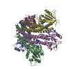
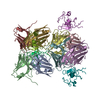
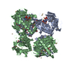
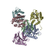
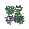
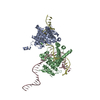
Crystal structure of the cytosolic domain of the CorA magnesium channel from Escherichia coli in complex with magnesium
ComponentsMagnesium transport protein CorA
KeywordsTRANSPORT PROTEIN / Homopentamer Complex Transport Membrane
Function / homology
Function and homology information


nickel cation transmembrane transport / nickel cation transmembrane transporter activity / magnesium ion transport / cobalt ion transport / cobalt ion transmembrane transporter activity / magnesium ion transmembrane transporter activity / plasma membrane
Similarity search - Function
Magnesium/cobalt transport protein CorA / CorA, cytoplasmic domain / CorA, transmembrane region / Mg2+ transporter protein, CorA-like/Zinc transport protein ZntB / CorA-like Mg2+ transporter protein
Similarity search - Domain/homology
Magnesium transport protein CorA
Similarity search - Component
Biological speciesEscherichia coli (E. coli)
MethodX-RAY DIFFRACTION / SYNCHROTRON / MOLECULAR REPLACEMENT / Resolution: 2.8 Å
AuthorsLerche, M. / Sandhu, H. / Flockner, L. / Hogbom, M. / Rapp, M.
Funding support Sweden, 4items
OrganizationGrant numberCountry
Swedish Research Council Sweden
Carl Tryggers stiftelse Sweden
Knut and Alice Wallenberg Foundation Sweden
Swedish Foundation for Strategic Research Sweden
CitationJournal: Structure / Year: 2017
Title: Structure and Cooperativity of the Cytosolic Domain of the CorA Mg(2+) Channel from Escherichia coli.
Authors: Lerche, M. / Sandhu, H. / Flockner, L. / Hogbom, M. / Rapp, M.
History
DepositionFeb 20, 2017Deposition site: PDBE / Processing site: PDBE
Revision 1.0Jul 12, 2017Provider: repository / Type: Initial release
Revision 1.1Aug 9, 2017Group: Author supporting evidence / Database references / Category: citation / pdbx_audit_support
Item: _citation.journal_volume / _citation.page_first ..._citation.journal_volume / _citation.page_first / _citation.page_last / _pdbx_audit_support.funding_organization
Revision 1.2Sep 13, 2017Group: Author supporting evidence / Category: pdbx_audit_support / Item: _pdbx_audit_support.funding_organization
Revision 1.3May 8, 2024Group: Data collection / Database references / Derived calculations
Category: chem_comp_atom / chem_comp_bond ...chem_comp_atom / chem_comp_bond / database_2 / pdbx_struct_conn_angle / struct_conn
Item: _database_2.pdbx_DOI / _database_2.pdbx_database_accession ..._database_2.pdbx_DOI / _database_2.pdbx_database_accession / _pdbx_struct_conn_angle.ptnr1_auth_asym_id / _pdbx_struct_conn_angle.ptnr1_auth_comp_id / _pdbx_struct_conn_angle.ptnr1_auth_seq_id / _pdbx_struct_conn_angle.ptnr1_label_asym_id / _pdbx_struct_conn_angle.ptnr1_label_atom_id / _pdbx_struct_conn_angle.ptnr1_label_comp_id / _pdbx_struct_conn_angle.ptnr1_label_seq_id / _pdbx_struct_conn_angle.ptnr3_auth_asym_id / _pdbx_struct_conn_angle.ptnr3_auth_comp_id / _pdbx_struct_conn_angle.ptnr3_auth_seq_id / _pdbx_struct_conn_angle.ptnr3_label_asym_id / _pdbx_struct_conn_angle.ptnr3_label_atom_id / _pdbx_struct_conn_angle.ptnr3_label_comp_id / _pdbx_struct_conn_angle.ptnr3_label_seq_id / _pdbx_struct_conn_angle.value / _struct_conn.pdbx_dist_value / _struct_conn.ptnr1_auth_asym_id / _struct_conn.ptnr1_auth_comp_id / _struct_conn.ptnr1_auth_seq_id / _struct_conn.ptnr1_label_asym_id / _struct_conn.ptnr1_label_atom_id / _struct_conn.ptnr1_label_comp_id / _struct_conn.ptnr1_label_seq_id / _struct_conn.ptnr2_auth_asym_id / _struct_conn.ptnr2_auth_comp_id / _struct_conn.ptnr2_auth_seq_id / _struct_conn.ptnr2_label_asym_id / _struct_conn.ptnr2_label_atom_id / _struct_conn.ptnr2_label_comp_id / _struct_conn.ptnr2_label_seq_id

-
Structure visualization

Structure viewerMolecule:
MolmilJmol/JSmol

Downloads & links

-
Assembly

Deposited unit
A: Magnesium transport protein CorA
B: Magnesium transport protein CorA
C: Magnesium transport protein CorA
D: Magnesium transport protein CorA
E: Magnesium transport protein CorA
hetero molecules


Theoretical massNumber of molelcules
Total (without water)149,88315
Polymers149,3535
Non-polymers53110
Water2,072115
1


  • Idetical with deposited unit
  • defined by author&software
TypeNameSymmetry operationNumber
identity operation1_555x,y,z1
Buried area17230 Å2
ΔGint-106 kcal/mol
Surface area52860 Å2
MethodPISA
Unit cell
Length a, b, c (Å)73.313, 117.453, 91.114
Angle α, β, γ (deg.)90.00, 103.27, 90.00
Int Tables number4
Space group name H-MP1211
Noncrystallographic symmetry (NCS)NCS domain:
IDEns-IDDetails
11C
21D
12C
22A
13C
23B
14C
24E
15D
25A
16D
26B
17D
27E
18A
28B
19A
29E
110B
210E

NCS domain segments:
Dom-IDComponent-IDEns-IDRefine codeAuth asym-IDAuth seq-ID
1010C1 - 258
2010D1 - 258
1020C1 - 258
2020A1 - 258
1030C1 - 258
2030B1 - 258
1040C1 - 258
2040E1 - 258
1050D1 - 258
2050A1 - 258
1060D1 - 258
2060B1 - 258
1070D1 - 258
2070E1 - 258
1080A1 - 258
2080B1 - 258
1090A1 - 258
2090E1 - 258
10100B1 - 258
20100E1 - 258

NCS ensembles :
ID
1
2
3
4
5
6
7
8
9
10

-
Components

#1: Protein
Magnesium transport protein CorA


Mass: 29870.543 Da / Num. of mol.: 5
Source method: isolated from a genetically manipulated source
Source: (gene. exp.) Escherichia coli (E. coli) / Gene: corA, b3816, JW3789 / Production host: Escherichia coli (E. coli) / References: UniProt: P0ABI4
#2: Chemical
ChemComp-MG / MAGNESIUM ION


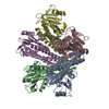
Mass: 24.305 Da / Num. of mol.: 7 / Source method: obtained synthetically / Formula: Mg
#3: Chemical ChemComp-PG0 / 2-(2-METHOXYETHOXY)ETHANOL / PEG 6000


Mass: 120.147 Da / Num. of mol.: 3 / Source method: obtained synthetically / Formula: C5H12O3 / Comment: inhibitor, precipitant*YM
#4: Water ChemComp-HOH / water


Mass: 18.015 Da / Num. of mol.: 115 / Source method: isolated from a natural source / Formula: H2O

-
Experimental details

-
Experiment

ExperimentMethod: X-RAY DIFFRACTION / Number of used crystals: 1

-
Sample preparation

CrystalDensity Matthews: 2.56 Å3/Da / Density % sol: 51.89 %
Crystal growTemperature: 291 K / Method: vapor diffusion, hanging drop
Details: 20 mM MgCl2; 100 mM Glycine pH8.7; 8-18% PEG400; 0,5-2% TritonX-114.

-
Data collection

DiffractionMean temperature: 100 K
Diffraction sourceSource: SYNCHROTRON / Site: Diamond / Beamline: I02 / Wavelength: 0.9795 Å
DetectorType: ADSC QUANTUM 315 / Detector: CCD / Date: Nov 29, 2010
RadiationProtocol: SINGLE WAVELENGTH / Monochromatic (M) / Laue (L): M / Scattering type: x-ray
Radiation wavelengthWavelength: 0.9795 Å / Relative weight: 1
ReflectionResolution: 2.8→29.84 Å / Num. obs: 36878 / % possible obs: 99 % / Redundancy: 6.2 % / CC1/2: 0.995 / Rmerge(I) obs: 0.129 / Net I/σ(I): 13
Reflection shellResolution: 2.8→2.95 Å / Redundancy: 5.8 % / Rmerge(I) obs: 0.836 / Mean I/σ(I) obs: 2.4 / CC1/2: 0.746 / % possible all: 93.5

-
Processing

Software
NameVersionClassification
REFMAC5.8.0073refinement
XDSdata reduction
SCALAdata scaling
PHASERphasing
RefinementMethod to determine structure: MOLECULAR REPLACEMENT / Resolution: 2.8→29.84 Å / Cor.coef. Fo:Fc: 0.949 / Cor.coef. Fo:Fc free: 0.928 / SU B: 36.054 / SU ML: 0.313 / Cross valid method: THROUGHOUT / ESU R Free: 0.37 / Stereochemistry target values: MAXIMUM LIKELIHOOD / Details: HYDROGENS HAVE BEEN ADDED IN THE RIDING POSITIONS
RfactorNum. reflection% reflectionSelection details
Rfree0.23591 1869 5.1 %RANDOM
Rwork0.19461 ---
obs0.19672 34990 98.92 %-
Solvent computationIon probe radii: 0.8 Å / Shrinkage radii: 0.8 Å / VDW probe radii: 1.2 Å / Solvent model: MASK
Displacement parametersBiso mean: 65.456 Å2
Baniso -1Baniso -2Baniso -3
1--1.89 Å20 Å21.35 Å2
2---2.07 Å20 Å2
3---2.99 Å2
Refinement stepCycle: 1 / Resolution: 2.8→29.84 Å
ProteinNucleic acidLigandSolventTotal
Num. atoms10387 0 31 115 10533
Refine LS restraints
Refine-IDTypeDev idealDev ideal targetNumber
X-RAY DIFFRACTIONr_bond_refined_d0.0110.01910598
X-RAY DIFFRACTIONr_bond_other_d0.0050.0210066
X-RAY DIFFRACTIONr_angle_refined_deg1.3711.96814287
X-RAY DIFFRACTIONr_angle_other_deg1.03323034
X-RAY DIFFRACTIONr_dihedral_angle_1_deg5.95351278
X-RAY DIFFRACTIONr_dihedral_angle_2_deg39.70424.328603
X-RAY DIFFRACTIONr_dihedral_angle_3_deg19.136151915
X-RAY DIFFRACTIONr_dihedral_angle_4_deg19.10715108
X-RAY DIFFRACTIONr_chiral_restr0.070.21582
X-RAY DIFFRACTIONr_gen_planes_refined0.0060.0212145
X-RAY DIFFRACTIONr_gen_planes_other0.0050.022521
X-RAY DIFFRACTIONr_nbd_refined
X-RAY DIFFRACTIONr_nbd_other
X-RAY DIFFRACTIONr_nbtor_refined
X-RAY DIFFRACTIONr_nbtor_other
X-RAY DIFFRACTIONr_xyhbond_nbd_refined
X-RAY DIFFRACTIONr_xyhbond_nbd_other
X-RAY DIFFRACTIONr_metal_ion_refined
X-RAY DIFFRACTIONr_metal_ion_other
X-RAY DIFFRACTIONr_symmetry_vdw_refined
X-RAY DIFFRACTIONr_symmetry_vdw_other
X-RAY DIFFRACTIONr_symmetry_hbond_refined
X-RAY DIFFRACTIONr_symmetry_hbond_other
X-RAY DIFFRACTIONr_symmetry_metal_ion_refined
X-RAY DIFFRACTIONr_symmetry_metal_ion_other
X-RAY DIFFRACTIONr_mcbond_it3.0864.355133
X-RAY DIFFRACTIONr_mcbond_other3.0874.355132
X-RAY DIFFRACTIONr_mcangle_it5.0526.5246404
X-RAY DIFFRACTIONr_mcangle_other5.0526.5246405
X-RAY DIFFRACTIONr_scbond_it4.1874.8445465
X-RAY DIFFRACTIONr_scbond_other4.1874.8455466
X-RAY DIFFRACTIONr_scangle_it
X-RAY DIFFRACTIONr_scangle_other6.8197.077884
X-RAY DIFFRACTIONr_long_range_B_refined9.42233.68611550
X-RAY DIFFRACTIONr_long_range_B_other9.42233.69311551
X-RAY DIFFRACTIONr_rigid_bond_restr
X-RAY DIFFRACTIONr_sphericity_free
X-RAY DIFFRACTIONr_sphericity_bonded
Refine LS restraints NCS

Refine-ID: X-RAY DIFFRACTION / Type: interatomic distance / Weight position: 0.05

Ens-IDDom-IDAuth asym-IDNumberRms dev position (Å)
11C158830.07
12D158830.07
21C157650.07
22A157650.07
31C160410.05
32B160410.05
41C158960.06
42E158960.06
51D160790.06
52A160790.06
61D160610.06
62B160610.06
71D161250.05
72E161250.05
81A158860.07
82B158860.07
91A160630.06
92E160630.06
101B160010.06
102E160010.06
LS refinement shellResolution: 2.795→2.868 Å / Total num. of bins used: 20
RfactorNum. reflection% reflection
Rfree0.384 111 -
Rwork0.374 2252 -
obs--86.91 %
Refinement TLS params.

Method: refined / Refine-ID: X-RAY DIFFRACTION

IDL112)L122)L132)L222)L232)L332)S11 (Å °)S12 (Å °)S13 (Å °)S21 (Å °)S22 (Å °)S23 (Å °)S31 (Å °)S32 (Å °)S33 (Å °)T112)T122)T132)T222)T232)T332)Origin x (Å)Origin y (Å)Origin z (Å)
12.05110.42281.05331.88940.33771.84120.13260.03-0.109-0.19590.0160.28430.2637-0.1201-0.14850.0711-0.0228-0.0550.0182-0.00930.2023-49.09630.0009-9.2676
21.95540.70630.06332.01950.00881.65580.1157-0.2347-0.29590.3566-0.01140.07350.4188-0.3212-0.10430.1733-0.07540.01040.09450.07820.1451-49.1476-4.016722.639
31.9994-0.77490.63471.5040.00081.89960.07770.1872-0.0591-0.3587-0.06630.1261-0.3044-0.202-0.01150.13730.0486-0.01130.060.00460.1061-48.732431.228-14.8641
41.8008-1.0664-0.62142.14790.51261.20130.1491-0.00170.1541-0.06120.04360.0906-0.2326-0.1874-0.19270.04750.0370.04460.03960.030.1295-49.08946.561812.9983
51.80880.1476-1.45631.2947-0.37052.27120.0864-0.1261-0.07470.40240.10420.1528-0.1355-0.1102-0.19060.12780.03050.04340.05990.060.0695-49.435824.621636.3318
Refinement TLS group
IDRefine-IDRefine TLS-IDAuth asym-IDAuth seq-ID
1X-RAY DIFFRACTION1C1 - 257
2X-RAY DIFFRACTION2D1 - 257
3X-RAY DIFFRACTION3A1 - 257
4X-RAY DIFFRACTION4B1 - 257
5X-RAY DIFFRACTION5E1 - 257

+
About Yorodumi

-
News

-
Feb 9, 2022. New format data for meta-information of EMDB entries

New format data for meta-information of EMDB entries

  • Version 3 of the EMDB header file is now the official format.
  • The previous official version 1.9 will be removed from the archive.

Related info.:EMDB header

External links:wwPDB to switch to version 3 of the EMDB data model

-
Aug 12, 2020. Covid-19 info

Covid-19 info

URL: https://pdbjlvh1.pdbj.org/emnavi/covid19.php

New page: Covid-19 featured information page in EM Navigator.

Related info.:Covid-19 info / Mar 5, 2020. Novel coronavirus structure data

+
Mar 5, 2020. Novel coronavirus structure data

Novel coronavirus structure data

Related info.:Yorodumi Speices / Aug 12, 2020. Covid-19 info

External links:COVID-19 featured content - PDBj / Molecule of the Month (242):Coronavirus Proteases

+
Jan 31, 2019. EMDB accession codes are about to change! (news from PDBe EMDB page)

EMDB accession codes are about to change! (news from PDBe EMDB page)

  • The allocation of 4 digits for EMDB accession codes will soon come to an end. Whilst these codes will remain in use, new EMDB accession codes will include an additional digit and will expand incrementally as the available range of codes is exhausted. The current 4-digit format prefixed with “EMD-” (i.e. EMD-XXXX) will advance to a 5-digit format (i.e. EMD-XXXXX), and so on. It is currently estimated that the 4-digit codes will be depleted around Spring 2019, at which point the 5-digit format will come into force.
  • The EM Navigator/Yorodumi systems omit the EMD- prefix.

Related info.:Q: What is EMD? / ID/Accession-code notation in Yorodumi/EM Navigator

External links:EMDB Accession Codes are Changing Soon! / Contact to PDBj

+
Jul 12, 2017. Major update of PDB

Major update of PDB

  • wwPDB released updated PDB data conforming to the new PDBx/mmCIF dictionary.
  • This is a major update changing the version number from 4 to 5, and with Remediation, in which all the entries are updated.
  • In this update, many items about electron microscopy experimental information are reorganized (e.g. em_software).
  • Now, EM Navigator and Yorodumi are based on the updated data.

External links:wwPDB Remediation / Enriched Model Files Conforming to OneDep Data Standards Now Available in the PDB FTP Archive

-
Yorodumi

Thousand views of thousand structures

  • Yorodumi is a browser for structure data from EMDB, PDB, SASBDB, etc.
  • This page is also the successor to EM Navigator detail page, and also detail information page/front-end page for Omokage search.
  • The word "yorodu" (or yorozu) is an old Japanese word meaning "ten thousand". "mi" (miru) is to see.

Related info.:EMDB / PDB / SASBDB / Comparison of 3 databanks / Yorodumi Search / Aug 31, 2016. New EM Navigator & Yorodumi / Yorodumi Papers / Jmol/JSmol / Function and homology information / Changes in new EM Navigator and Yorodumi

Read more