 Movie
Movie Controller

[English] 日本語
 Yorodumi
- PDB-5n2v: Changes in conformational equilibria regulate the activity of the... -
+ Open data

- Basic information
| Entry | Database: PDB / ID: 5n2v | |||||||||
|---|---|---|---|---|---|---|---|---|---|---|
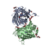
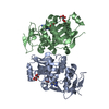
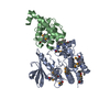
| Title | Changes in conformational equilibria regulate the activity of the Dcp2 decapping enzyme | |||||||||
 Components Components |
| |||||||||
 Keywords Keywords | RNA BINDING PROTEIN | |||||||||
| Function / homology |  Function and homology information Function and homology informationmRNA phosphatase activator activity / RNA decapping complex / mRNA methylguanosine-cap decapping / 5'-(N(7)-methyl 5'-triphosphoguanosine)-[mRNA] diphosphatase activity / 5'-(N(7)-methylguanosine 5'-triphospho)-[mRNA] hydrolase activity / Hydrolases / deadenylation-dependent decapping of nuclear-transcribed mRNA / mRNA cap binding / nuclear-transcribed mRNA catabolic process, nonsense-mediated decay / nuclear-transcribed mRNA catabolic process ...mRNA phosphatase activator activity / RNA decapping complex / mRNA methylguanosine-cap decapping / 5'-(N(7)-methyl 5'-triphosphoguanosine)-[mRNA] diphosphatase activity / 5'-(N(7)-methylguanosine 5'-triphospho)-[mRNA] hydrolase activity / Hydrolases / deadenylation-dependent decapping of nuclear-transcribed mRNA / mRNA cap binding / nuclear-transcribed mRNA catabolic process, nonsense-mediated decay / nuclear-transcribed mRNA catabolic process / P-body / mRNA processing / cytoplasmic stress granule / manganese ion binding / single-stranded RNA binding / magnesium ion binding / RNA binding / ATP binding / nucleus / cytosol / cytoplasm Similarity search - Function | |||||||||
| Biological species |  | |||||||||
| Method |  X-RAY DIFFRACTION / SYNCHROTRON / MOLECULAR REPLACEMENT / Resolution: 3.1 Å X-RAY DIFFRACTION / SYNCHROTRON / MOLECULAR REPLACEMENT / Resolution: 3.1 Å | |||||||||
 Authors Authors | Holdermann, I. / Sprangers, R. | |||||||||
| Funding support | 1items
| |||||||||
 Citation Citation | Journal: Proc. Natl. Acad. Sci. U.S.A. / Year: 2017 Title: Changes in conformational equilibria regulate the activity of the Dcp2 decapping enzyme. Authors: Wurm, J.P. / Holdermann, I. / Overbeck, J.H. / Mayer, P.H.O. / Sprangers, R. | |||||||||
| History |
|
- Structure visualization
| Structure viewer | Molecule: MolmilJmol/JSmol |
|---|
- Downloads & links
-Download
| PDBx/mmCIF format | 5n2v.cif.gz | 166.3 KB | Display | PDBx/mmCIF format |
|---|---|---|---|---|
| PDB format | pdb5n2v.ent.gz | 129.4 KB | Display | PDB format |
| PDBx/mmJSON format | 5n2v.json.gz | Tree view | PDBx/mmJSON format | |
| Others |  Other downloads Other downloads |
-Validation report
| Arichive directory | https://data.pdbj.org/pub/pdb/validation_reports/n2/5n2vftp://data.pdbj.org/pub/pdb/validation_reports/n2/5n2v | HTTPS FTP |
|---|
-Related structure data
| Related structure data | 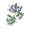 5j3tS S: Starting model for refinement |
|---|---|
| Similar structure data |


-Links
 PDBj
PDBj


- Assembly
| Deposited unit | 
| ||||||||
|---|---|---|---|---|---|---|---|---|---|
| 1 | 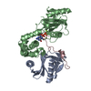
| ||||||||
| 2 | 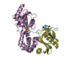
| ||||||||
| Unit cell |
|
-Components
| #1: Protein | Mass: 15130.271 Da / Num. of mol.: 2 Source method: isolated from a genetically manipulated source Source: (gene. exp.) Gene: dcp1, SPBC3B9.21 / Production host:  #2: Protein | Mass: 28689.143 Da / Num. of mol.: 2 Source method: isolated from a genetically manipulated source Source: (gene. exp.) Gene: dcp2, SPAC19A8.12 / Production host: #3: Protein/peptide | Mass: 2742.089 Da / Num. of mol.: 2 / Source method: obtained synthetically / Source: (synth.) #4: Chemical | ChemComp-MG /   Mass: 24.305 Da / Num. of mol.: 6 / Source method: obtained synthetically / Formula: Mg Mass: 24.305 Da / Num. of mol.: 6 / Source method: obtained synthetically / Formula: Mg#5: Chemical |   Mass: 459.243 Da / Num. of mol.: 2 / Source method: obtained synthetically / Formula: C11H19N5O11P2 Mass: 459.243 Da / Num. of mol.: 2 / Source method: obtained synthetically / Formula: C11H19N5O11P2 |
|---|
-Experimental details
-Experiment
| Experiment | Method: X-RAY DIFFRACTION / Number of used crystals: 1 |
|---|
- Sample preparation
| Crystal | Density Matthews: 2.29 Å3/Da / Density % sol: 46.28 % |
|---|---|
| Crystal grow | Temperature: 293 K / Method: vapor diffusion, sitting drop / pH: 7 Details: 41% M1K3350 Morpheus Mix 4, MOPS pH 7.0, 0.06 M MgCl2/CaCl2 |
-Data collection
| Diffraction | Mean temperature: 100 K |
|---|---|
| Diffraction source | Source: SYNCHROTRON / Site: SLS  / Beamline: X10SA / Wavelength: 0.99 Å / Beamline: X10SA / Wavelength: 0.99 Å |
| Detector | Type: PSI PILATUS 6M / Detector: PIXEL / Date: Oct 2, 2016 |
| Radiation | Protocol: SINGLE WAVELENGTH / Monochromatic (M) / Laue (L): M / Scattering type: x-ray |
| Radiation wavelength | Wavelength: 0.99 Å / Relative weight: 1 |
| Reflection | Resolution: 3.1→48.3 Å / Num. obs: 15440 / % possible obs: 99.9 % / Redundancy: 3.4 % / CC1/2: 0.95 / Rmerge(I) obs: 0.24 / Net I/σ(I): 4.3 |
| Reflection shell | Resolution: 3.1→3.27 Å / Redundancy: 3.4 % / Rmerge(I) obs: 0.73 / Mean I/σ(I) obs: 1.8 / CC1/2: 0.64 / % possible all: 100 |
- Processing
| Software |
| |||||||||||||||||||||||||||||||||||||||||||||||||
|---|---|---|---|---|---|---|---|---|---|---|---|---|---|---|---|---|---|---|---|---|---|---|---|---|---|---|---|---|---|---|---|---|---|---|---|---|---|---|---|---|---|---|---|---|---|---|---|---|---|---|
| Refinement | Method to determine structure: MOLECULAR REPLACEMENT Starting model: 5j3t Resolution: 3.1→48.289 Å / SU ML: 0.43 / Cross valid method: FREE R-VALUE / σ(F): 1.34 / Phase error: 29.97
| |||||||||||||||||||||||||||||||||||||||||||||||||
| Solvent computation | Shrinkage radii: 0.9 Å / VDW probe radii: 1.11 Å | |||||||||||||||||||||||||||||||||||||||||||||||||
| Refinement step | Cycle: LAST / Resolution: 3.1→48.289 Å
| |||||||||||||||||||||||||||||||||||||||||||||||||
| Refine LS restraints |
| |||||||||||||||||||||||||||||||||||||||||||||||||
| LS refinement shell |
|
