 ムービー
ムービー コントローラー


+ データを開く

- 基本情報
基本情報
| 登録情報 | データベース: PDB / ID: 5mz6 | ||||||||||||||||||||||||||||||||||||||||||||||||||||||||||||
|---|---|---|---|---|---|---|---|---|---|---|---|---|---|---|---|---|---|---|---|---|---|---|---|---|---|---|---|---|---|---|---|---|---|---|---|---|---|---|---|---|---|---|---|---|---|---|---|---|---|---|---|---|---|---|---|---|---|---|---|---|---|
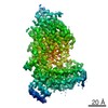
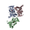
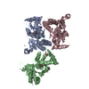
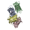
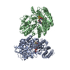
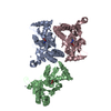
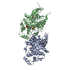
| タイトル | Cryo-EM structure of a Separase-Securin complex from Caenorhabditis elegans at 3.8 A resolution | ||||||||||||||||||||||||||||||||||||||||||||||||||||||||||||
 要素 要素 |
| ||||||||||||||||||||||||||||||||||||||||||||||||||||||||||||
 キーワード キーワード | CELL CYCLE / caspase / cohesin / cleavage | ||||||||||||||||||||||||||||||||||||||||||||||||||||||||||||
| 機能・相同性 |  機能・相同性情報 機能・相同性情報Separation of Sister Chromatids / separase-securin complex / eggshell formation / metaphase plate / regulation of nematode larval development / separase / multicellular organismal reproductive process / maintenance of meiotic sister chromatid cohesion / cortical granule exocytosis / meiotic chromosome separation ...Separation of Sister Chromatids / separase-securin complex / eggshell formation / metaphase plate / regulation of nematode larval development / separase / multicellular organismal reproductive process / maintenance of meiotic sister chromatid cohesion / cortical granule exocytosis / meiotic chromosome separation / maintenance of mitotic sister chromatid cohesion / polarity specification of anterior/posterior axis / polar body extrusion after meiotic divisions / cortical granule / regulation of centriole-centriole cohesion / mitotic sister chromatid separation / regulation of exocytosis / meiotic spindle / centrosome localization / regulation of locomotion / cleavage furrow / mitotic cytokinesis / centrosome duplication / condensed chromosome / condensed nuclear chromosome / spindle microtubule / protein processing / spindle / mitotic spindle / intracellular protein localization / nuclear envelope / chromosome / midbody / protease binding / cell cortex / protein stabilization / cysteine-type endopeptidase activity / ubiquitin protein ligase binding / centrosome / nucleus / cytoplasm 類似検索 - 分子機能 | ||||||||||||||||||||||||||||||||||||||||||||||||||||||||||||
| 生物種 |  | ||||||||||||||||||||||||||||||||||||||||||||||||||||||||||||
| 手法 | 電子顕微鏡法 / 単粒子再構成法 / クライオ電子顕微鏡法 / 解像度: 3.8 Å | ||||||||||||||||||||||||||||||||||||||||||||||||||||||||||||
 データ登録者 データ登録者 | Boland, A. / Martin, T.G. / Zhang, Z. / Yang, J. / Bai, X.C. / Chang, L. / Scheres, S.H.W. / Barford, D. | ||||||||||||||||||||||||||||||||||||||||||||||||||||||||||||
 引用 引用 | ジャーナル: Nat Struct Mol Biol / 年: 2017 タイトル: Cryo-EM structure of a metazoan separase-securin complex at near-atomic resolution. 著者: Andreas Boland / Thomas G Martin / Ziguo Zhang / Jing Yang / Xiao-Chen Bai / Leifu Chang / Sjors H W Scheres / David Barford /  要旨: Separase is a caspase-family protease that initiates chromatid segregation by cleaving the kleisin subunits (Scc1 and Rec8) of cohesin, and regulates centrosome duplication and mitotic spindle ...Separase is a caspase-family protease that initiates chromatid segregation by cleaving the kleisin subunits (Scc1 and Rec8) of cohesin, and regulates centrosome duplication and mitotic spindle function through cleavage of kendrin and Slk19. To understand the mechanisms of securin regulation of separase, we used single-particle cryo-electron microscopy (cryo-EM) to determine a near-atomic-resolution structure of the Caenorhabditis elegans separase-securin complex. Separase adopts a triangular-shaped bilobal architecture comprising an N-terminal tetratricopeptide repeat (TPR)-like α-solenoid domain docked onto the conserved C-terminal protease domain. Securin engages separase in an extended antiparallel conformation, interacting with both lobes. It inhibits separase by interacting with the catalytic site through a pseudosubstrate mechanism, thus revealing that in the inhibited separase-securin complex, the catalytic site adopts a conformation compatible with substrate binding. Securin is protected from cleavage because an aliphatic side chain at the P1 position represses protease activity by disrupting the organization of catalytic site residues. | ||||||||||||||||||||||||||||||||||||||||||||||||||||||||||||
| 履歴 |
|
- 構造の表示
構造の表示
| ムービー |
ムービービューア |
|---|---|
| 構造ビューア | 分子: MolmilJmol/JSmol |
- ダウンロードとリンク
-ダウンロード
| PDBx/mmCIF形式 | 5mz6.cif.gz | 208.8 KB | 表示 | PDBx/mmCIF形式 |
|---|---|---|---|---|
| PDB形式 | pdb5mz6.ent.gz | 156.8 KB | 表示 | PDB形式 |
| PDBx/mmJSON形式 | 5mz6.json.gz | ツリー表示 | PDBx/mmJSON形式 | |
| その他 |  その他のダウンロード その他のダウンロード |
-検証レポート
| 文書・要旨 | 5mz6_validation.pdf.gz | 704.7 KB | 表示 | wwPDB検証レポート |
|---|---|---|---|---|
| 文書・詳細版 | 5mz6_full_validation.pdf.gz | 717.5 KB | 表示 | |
| XML形式データ | 5mz6_validation.xml.gz | 33.6 KB | 表示 | |
| CIF形式データ | 5mz6_validation.cif.gz | 51.6 KB | 表示 | |
| アーカイブディレクトリ | https://data.pdbj.org/pub/pdb/validation_reports/mz/5mz6ftp://data.pdbj.org/pub/pdb/validation_reports/mz/5mz6 | HTTPS FTP |
-関連構造データ


-リンク
 PDBj
PDBj
- 集合体
集合体
| 登録構造単位 | 
|
|---|---|
| 1 |
|
-要素
| #1: タンパク質 | 分子量: 144315.984 Da / 分子数: 1 / 由来タイプ: 組換発現 由来: (組換発現) 遺伝子: sep-1, CELE_Y47G6A.12, Y47G6A.12 / 発現宿主:  Trichoplusia ni (イラクサキンウワバ) / 参照: UniProt: G5ED39 Trichoplusia ni (イラクサキンウワバ) / 参照: UniProt: G5ED39 |
|---|---|
| #2: タンパク質 | 分子量: 27027.646 Da / 分子数: 1 / 由来タイプ: 組換発現 由来: (組換発現) 遺伝子: ify-1, C27A2.3, CELE_C27A2.3 / 発現宿主: Trichoplusia ni (イラクサキンウワバ) / 参照: UniProt: Q18235 |
| Has protein modification | Y |
-実験情報
-実験
| 実験 | 手法: 電子顕微鏡法 |
|---|---|
| EM実験 | 試料の集合状態: PARTICLE / 3次元再構成法: 単粒子再構成法 |
- 試料調製
試料調製
| 構成要素 | 名称: Caenorhabditis elegans Separase-Securin complex / タイプ: COMPLEX 詳細: C. elegans Separase-Securin complex at 3.8 Angstrom resolution refined with Relion. Entity ID: all / 由来: RECOMBINANT |
|---|---|
| 分子量 | 値: 0.17 MDa / 実験値: NO |
| 由来(天然) | 生物種: |
| 由来(組換発現) | 生物種: Trichoplusia ni (イラクサキンウワバ) |
| 緩衝液 | pH: 7.8 |
| 試料 | 濃度: 0.02 mg/ml / 包埋: NO / シャドウイング: NO / 染色: NO / 凍結: YES / 詳細: Monodisperse sample |
| 試料支持 | 詳細: Glow discharging was performed before applying graphene oxide solution onto the grid. Grid was washed three times, dried and sample was applied. For detailed information see: https://figshare. ...詳細: Glow discharging was performed before applying graphene oxide solution onto the grid. Grid was washed three times, dried and sample was applied. For detailed information see: https://figshare.com/articles/Graphene_Oxide_Grid_Preparation/3178669 グリッドの材料: GOLD / グリッドのサイズ: 300 divisions/in. / グリッドのタイプ: Quantifoil R1.2/1.3 |
| 急速凍結 | 凍結剤: ETHANE / 湿度: 100 % / 凍結前の試料温度: 277 K / 詳細: Custom Manual Plunger |
- 電子顕微鏡撮影
| 実験機器 |  モデル: Titan Krios / 画像提供: FEI Company |
|---|---|
| 顕微鏡 | モデル: FEI TITAN KRIOS |
| 電子銃 | 電子線源:  FIELD EMISSION GUN / 加速電圧: 300 kV / 照射モード: FLOOD BEAM FIELD EMISSION GUN / 加速電圧: 300 kV / 照射モード: FLOOD BEAM |
| 電子レンズ | モード: BRIGHT FIELD / Cs: 2.7 mm / C2レンズ絞り径: 70 µm / アライメント法: COMA FREE |
| 試料ホルダ | 凍結剤: NITROGEN 試料ホルダーモデル: FEI TITAN KRIOS AUTOGRID HOLDER |
| 撮影 | 平均露光時間: 0.8 sec. / 電子線照射量: 1.25 e/Å2 / 検出モード: SUPER-RESOLUTION フィルム・検出器のモデル: GATAN K2 SUMMIT (4k x 4k) |
| 電子光学装置 | エネルギーフィルター名称: GIF / エネルギーフィルター 上限: 20 eV / エネルギーフィルター 下限: -20 eV |
| 画像スキャン | 横: 3710 / 縦: 3710 |
- 解析
解析
| ソフトウェア | 名称: PHENIX / バージョン: 1.11.1_2575: / 分類: 精密化 | ||||||||||||||||||||||||||||
|---|---|---|---|---|---|---|---|---|---|---|---|---|---|---|---|---|---|---|---|---|---|---|---|---|---|---|---|---|---|
| EMソフトウェア |
| ||||||||||||||||||||||||||||
| EM 3D crystal entity | ∠α: 90 ° / ∠β: 90 ° / ∠γ: 90 ° / A: 1 Å / B: 1 Å / C: 1 Å / 空間群名: 1 / 空間群番号: 1 | ||||||||||||||||||||||||||||
| CTF補正 | タイプ: PHASE FLIPPING AND AMPLITUDE CORRECTION | ||||||||||||||||||||||||||||
| 対称性 | 点対称性: C1 (非対称) | ||||||||||||||||||||||||||||
| 3次元再構成 | 解像度: 3.8 Å / 解像度の算出法: FSC 0.143 CUT-OFF / 粒子像の数: 103696 / 対称性のタイプ: POINT | ||||||||||||||||||||||||||||
| 原子モデル構築 | プロトコル: AB INITIO MODEL / 空間: REAL | ||||||||||||||||||||||||||||
| 拘束条件 |
|
