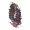
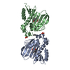
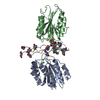
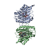
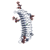
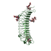
[English] 日本語
Yorodumi
- PDB-5mx0: Crystal structure of human fibromodulin -

+
Open data


ID or keywords:

Loading...

-
Basic information

Entry
Database: PDB / ID: 5mx0
TitleCrystal structure of human fibromodulin
ComponentsFibromodulin
KeywordsSTRUCTURAL PROTEIN / Leucine-rich repeat / extracellular matrix / collagen binding
Function / homology
Function and homology information


Defective CHST6 causes MCDC1 / Defective ST3GAL3 causes MCT12 and EIEE15 / transforming growth factor beta receptor complex assembly / keratan sulfate catabolic process / keratan sulfate biosynthetic process / Keratan sulfate degradation / Defective B4GALT1 causes B4GALT1-CDG (CDG-2d) / Keratan sulfate biosynthesis / collagen fibril organization / ECM proteoglycans ...Defective CHST6 causes MCDC1 / Defective ST3GAL3 causes MCT12 and EIEE15 / transforming growth factor beta receptor complex assembly / keratan sulfate catabolic process / keratan sulfate biosynthetic process / Keratan sulfate degradation / Defective B4GALT1 causes B4GALT1-CDG (CDG-2d) / Keratan sulfate biosynthesis / collagen fibril organization / ECM proteoglycans / lysosomal lumen / extracellular matrix / Golgi lumen / collagen-containing extracellular matrix / extracellular space / extracellular region
Similarity search - Function
Fibromodulin / Leucine rich repeat N-terminal domain / Leucine-rich repeat N-terminal domain / Leucine rich repeat N-terminal domain / Leucine rich repeat / Leucine-rich repeat, typical subtype / Leucine-rich repeats, typical (most populated) subfamily / Leucine-rich repeat profile. / Leucine-rich repeat / Leucine-rich repeat domain superfamily
Similarity search - Domain/homology
NICKEL (II) ION / Fibromodulin
Similarity search - Component
Biological speciesHomo sapiens (human)
MethodX-RAY DIFFRACTION / SYNCHROTRON / MOLECULAR REPLACEMENT / Resolution: 2.21 Å
AuthorsParacuellos, P. / Hohenester, E.
Funding support United Kingdom, 1items
OrganizationGrant numberCountry
Wellcome Trust101748/Z/13/Z United Kingdom
CitationJournal: Matrix Biol. / Year: 2017
Title: Structural and functional analysis of two small leucine-rich repeat proteoglycans, fibromodulin and chondroadherin.
Authors: Paracuellos, P. / Kalamajski, S. / Bonna, A. / Bihan, D. / Farndale, R.W. / Hohenester, E.
History
DepositionJan 20, 2017Deposition site: PDBE / Processing site: PDBE
Revision 1.0Mar 1, 2017Provider: repository / Type: Initial release
Revision 1.1Sep 27, 2017Group: Database references / Category: citation
Item: _citation.journal_volume / _citation.page_first / _citation.page_last
Revision 2.0Jul 29, 2020Group: Atomic model / Data collection ...Atomic model / Data collection / Derived calculations / Structure summary
Category: atom_site / atom_site_anisotrop ...atom_site / atom_site_anisotrop / chem_comp / entity / pdbx_branch_scheme / pdbx_chem_comp_identifier / pdbx_entity_branch / pdbx_entity_branch_descriptor / pdbx_entity_branch_link / pdbx_entity_branch_list / pdbx_entity_nonpoly / pdbx_nonpoly_scheme / pdbx_struct_assembly_gen / pdbx_struct_conn_angle / pdbx_struct_special_symmetry / struct_asym / struct_conn / struct_site / struct_site_gen
Item: _atom_site.B_iso_or_equiv / _atom_site.Cartn_x ..._atom_site.B_iso_or_equiv / _atom_site.Cartn_x / _atom_site.Cartn_y / _atom_site.Cartn_z / _atom_site.auth_asym_id / _atom_site.auth_atom_id / _atom_site.auth_comp_id / _atom_site.auth_seq_id / _atom_site.label_asym_id / _atom_site.label_atom_id / _atom_site.label_comp_id / _atom_site.label_entity_id / _atom_site.occupancy / _atom_site.type_symbol / _atom_site_anisotrop.U[1][1] / _atom_site_anisotrop.U[1][2] / _atom_site_anisotrop.U[1][3] / _atom_site_anisotrop.U[2][2] / _atom_site_anisotrop.U[2][3] / _atom_site_anisotrop.U[3][3] / _atom_site_anisotrop.pdbx_auth_asym_id / _atom_site_anisotrop.pdbx_auth_atom_id / _atom_site_anisotrop.pdbx_auth_comp_id / _atom_site_anisotrop.pdbx_auth_seq_id / _atom_site_anisotrop.pdbx_label_asym_id / _atom_site_anisotrop.pdbx_label_atom_id / _atom_site_anisotrop.pdbx_label_comp_id / _atom_site_anisotrop.type_symbol / _chem_comp.name / _chem_comp.type / _pdbx_struct_assembly_gen.asym_id_list / _pdbx_struct_conn_angle.ptnr1_auth_comp_id / _pdbx_struct_conn_angle.ptnr1_auth_seq_id / _pdbx_struct_conn_angle.ptnr1_label_asym_id / _pdbx_struct_conn_angle.ptnr1_label_atom_id / _pdbx_struct_conn_angle.ptnr1_label_comp_id / _pdbx_struct_conn_angle.ptnr1_label_seq_id / _pdbx_struct_conn_angle.ptnr2_label_asym_id / _pdbx_struct_conn_angle.ptnr2_symmetry / _pdbx_struct_conn_angle.ptnr3_auth_comp_id / _pdbx_struct_conn_angle.ptnr3_auth_seq_id / _pdbx_struct_conn_angle.ptnr3_label_asym_id / _pdbx_struct_conn_angle.ptnr3_label_atom_id / _pdbx_struct_conn_angle.ptnr3_label_comp_id / _pdbx_struct_conn_angle.ptnr3_label_seq_id / _pdbx_struct_conn_angle.value / _pdbx_struct_special_symmetry.label_asym_id / _struct_conn.conn_type_id / _struct_conn.id / _struct_conn.pdbx_dist_value / _struct_conn.pdbx_leaving_atom_flag / _struct_conn.pdbx_role / _struct_conn.ptnr1_auth_asym_id / _struct_conn.ptnr1_auth_comp_id / _struct_conn.ptnr1_auth_seq_id / _struct_conn.ptnr1_label_asym_id / _struct_conn.ptnr1_label_atom_id / _struct_conn.ptnr1_label_comp_id / _struct_conn.ptnr1_label_seq_id / _struct_conn.ptnr2_auth_asym_id / _struct_conn.ptnr2_auth_comp_id / _struct_conn.ptnr2_auth_seq_id / _struct_conn.ptnr2_label_asym_id / _struct_conn.ptnr2_label_atom_id / _struct_conn.ptnr2_label_comp_id / _struct_conn.ptnr2_symmetry
Description: Carbohydrate remediation / Provider: repository / Type: Remediation
Revision 2.1Jan 17, 2024Group: Data collection / Database references ...Data collection / Database references / Derived calculations / Refinement description / Structure summary
Category: chem_comp / chem_comp_atom ...chem_comp / chem_comp_atom / chem_comp_bond / database_2 / pdbx_initial_refinement_model / struct_conn
Item: _chem_comp.pdbx_synonyms / _database_2.pdbx_DOI ..._chem_comp.pdbx_synonyms / _database_2.pdbx_DOI / _database_2.pdbx_database_accession / _struct_conn.pdbx_leaving_atom_flag

-
Structure visualization

Structure viewerMolecule:
MolmilJmol/JSmol

Downloads & links

-
Assembly

Deposited unit
A: Fibromodulin
B: Fibromodulin
hetero molecules


Theoretical massNumber of molelcules
Total (without water)87,66914
Polymers81,6192
Non-polymers6,05012
Water10,196566
1
A: Fibromodulin
hetero molecules


Theoretical massNumber of molelcules
Total (without water)43,8357
Polymers40,8101
Non-polymers3,0256
Water181
TypeNameSymmetry operationNumber
identity operation1_555x,y,z1
MethodPISA
2
B: Fibromodulin
hetero molecules


Theoretical massNumber of molelcules
Total (without water)43,8357
Polymers40,8101
Non-polymers3,0256
Water181
TypeNameSymmetry operationNumber
identity operation1_555x,y,z1
MethodPISA
Unit cell
Length a, b, c (Å)108.110, 98.930, 111.340
Angle α, β, γ (deg.)90.00, 107.39, 90.00
Int Tables number5
Space group name H-MC121
Components on special symmetry positions
IDModelComponents
11A-874-

HOH

-
Components

#1: Protein Fibromodulin / FM / Collagen-binding 59 kDa protein / Keratan sulfate proteoglycan fibromodulin / KSPG fibromodulin


Mass: 40809.684 Da / Num. of mol.: 2
Mutation: Y38S, Y39S, Y42S, Y45S, Y47S, Y50S, Y53S, Y55S, Y63S, Y65S
Source method: isolated from a genetically manipulated source
Source: (gene. exp.) Homo sapiens (human) / Gene: FMOD, FM, SLRR2E / Plasmid: pCEP-Pu / Cell line (production host): HEK293 c18 / Production host: Homo sapiens (human) / References: UniProt: Q06828
#2: Polysaccharide
beta-D-mannopyranose-(1-4)-2-acetamido-2-deoxy-beta-D-glucopyranose-(1-4)-[alpha-L-fucopyranose-(1- ...beta-D-mannopyranose-(1-4)-2-acetamido-2-deoxy-beta-D-glucopyranose-(1-4)-[alpha-L-fucopyranose-(1-6)]2-acetamido-2-deoxy-beta-D-glucopyranose


Type: oligosaccharide / Mass: 732.682 Da / Num. of mol.: 8
Source method: isolated from a genetically manipulated source
DescriptorTypeProgram
DManpb1-4DGlcpNAcb1-4[LFucpa1-6]DGlcpNAcb1-Glycam Condensed SequenceGMML 1.0
WURCS=2.0/3,4,3/[a2122h-1b_1-5_2*NCC/3=O][a1122h-1b_1-5][a1221m-1a_1-5]/1-1-2-3/a4-b1_a6-d1_b4-c1WURCSPDB2Glycan 1.1.0
[]{[(4+1)][b-D-GlcpNAc]{[(4+1)][b-D-GlcpNAc]{[(4+1)][b-D-Manp]{}}[(6+1)][a-L-Fucp]{}}}LINUCSPDB-CARE
#3: Chemical ChemComp-CL / CHLORIDE ION


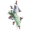
Mass: 35.453 Da / Num. of mol.: 2 / Source method: obtained synthetically / Formula: Cl
#4: Chemical ChemComp-NI / NICKEL (II) ION


Mass: 58.693 Da / Num. of mol.: 2 / Source method: obtained synthetically / Formula: Ni
#5: Water ChemComp-HOH / water


Mass: 18.015 Da / Num. of mol.: 566 / Source method: isolated from a natural source / Formula: H2O

-
Experimental details

-
Experiment

ExperimentMethod: X-RAY DIFFRACTION / Number of used crystals: 1

-
Sample preparation

CrystalDensity Matthews: 3.75 Å3/Da / Density % sol: 67.23 %
Crystal growTemperature: 295 K / Method: vapor diffusion, sitting drop
Details: 10 mg/ml protein + 26% (w/v) PEG 3350, 200 mM lithium sulphate monohydrate, 100 mM Tris-HCl (pH 8.5)

-
Data collection

DiffractionMean temperature: 100 K
Diffraction sourceSource: SYNCHROTRON / Site: Diamond / Beamline: I04-1 / Wavelength: 0.9282 Å
DetectorType: DECTRIS PILATUS3 6M / Detector: PIXEL / Date: Apr 28, 2016
RadiationProtocol: SINGLE WAVELENGTH / Monochromatic (M) / Laue (L): M / Scattering type: x-ray
Radiation wavelengthWavelength: 0.9282 Å / Relative weight: 1
ReflectionResolution: 2.21→65.913 Å / Num. obs: 55434 / % possible obs: 99 % / Redundancy: 6.8 % / CC1/2: 0.998 / Rmerge(I) obs: 0.111 / Rpim(I) all: 0.046 / Net I/σ(I): 9.5
Reflection shellResolution: 2.21→2.27 Å / Redundancy: 6.9 % / Rmerge(I) obs: 1.84 / Mean I/σ(I) obs: 1.1 / CC1/2: 0.523 / Rpim(I) all: 0.737 / % possible all: 99.1

-
Processing

Software
NameVersionClassification
PHENIX1.9_1692refinement
xia2data reduction
xia2data scaling
PHENIXphasing
RefinementMethod to determine structure: MOLECULAR REPLACEMENT
Starting model: 1XKU
Resolution: 2.21→65.913 Å / SU ML: 0.28 / Cross valid method: FREE R-VALUE / σ(F): 1.34 / Phase error: 27.93 / Stereochemistry target values: ML
RfactorNum. reflection% reflection
Rfree0.2162 2823 5.1 %
Rwork0.1793 --
obs0.1812 55367 98.81 %
Solvent computationShrinkage radii: 0.9 Å / VDW probe radii: 1.11 Å / Solvent model: FLAT BULK SOLVENT MODEL
Refinement stepCycle: LAST / Resolution: 2.21→65.913 Å
ProteinNucleic acidLigandSolventTotal
Num. atoms4881 0 396 566 5843
Refine LS restraints
Refine-IDTypeDev idealNumber
X-RAY DIFFRACTIONf_bond_d0.0035416
X-RAY DIFFRACTIONf_angle_d0.7597408
X-RAY DIFFRACTIONf_dihedral_angle_d11.4972108
X-RAY DIFFRACTIONf_chiral_restr0.03890
X-RAY DIFFRACTIONf_plane_restr0.003909
LS refinement shell
Resolution (Å)Rfactor RfreeNum. reflection RfreeRfactor RworkNum. reflection RworkRefine-ID% reflection obs (%)
2.21-2.24810.3181310.29722609X-RAY DIFFRACTION99
2.2481-2.2890.35111300.32542661X-RAY DIFFRACTION99
2.289-2.3330.33111370.28952553X-RAY DIFFRACTION99
2.333-2.38070.32481310.28932621X-RAY DIFFRACTION98
2.3807-2.43240.34121560.27662591X-RAY DIFFRACTION98
2.4324-2.4890.29781280.25992629X-RAY DIFFRACTION100
2.489-2.55130.2671520.24632638X-RAY DIFFRACTION100
2.5513-2.62020.27111520.22782643X-RAY DIFFRACTION100
2.6202-2.69730.25251240.22192640X-RAY DIFFRACTION100
2.6973-2.78440.30251450.2232631X-RAY DIFFRACTION100
2.7844-2.88390.26691540.21542589X-RAY DIFFRACTION99
2.8839-2.99940.26361340.22322635X-RAY DIFFRACTION98
2.9994-3.13590.2751330.22692579X-RAY DIFFRACTION97
3.1359-3.30120.25971350.21792675X-RAY DIFFRACTION99
3.3012-3.5080.20861570.18442641X-RAY DIFFRACTION100
3.508-3.77890.17631330.15612634X-RAY DIFFRACTION99
3.7789-4.15910.17461510.12412653X-RAY DIFFRACTION99
4.1591-4.76080.14491290.11242586X-RAY DIFFRACTION97
4.7608-5.99750.17351430.12922678X-RAY DIFFRACTION99
5.9975-65.94210.18751680.16412658X-RAY DIFFRACTION98
Refinement TLS params.Method: refined / Origin x: -41.2711 Å / Origin y: 19.1171 Å / Origin z: 132.5475 Å
111213212223313233
T0.3546 Å20.0076 Å2-0.036 Å2-0.3001 Å2-0.009 Å2--0.3275 Å2
L0.7149 °20.0881 °2-0.3375 °2-0.3527 °2-0.0733 °2--0.6785 °2
S0.1025 Å °-0.0115 Å °-0.0163 Å °0.009 Å °-0.0378 Å °-0.0048 Å °0.0363 Å °0.0005 Å °0.0077 Å °
Refinement TLS groupSelection details: all

+
About Yorodumi

-
News

-
Feb 9, 2022. New format data for meta-information of EMDB entries

New format data for meta-information of EMDB entries

  • Version 3 of the EMDB header file is now the official format.
  • The previous official version 1.9 will be removed from the archive.

Related info.:EMDB header

External links:wwPDB to switch to version 3 of the EMDB data model

-
Aug 12, 2020. Covid-19 info

Covid-19 info

URL: https://pdbjlvh1.pdbj.org/emnavi/covid19.php

New page: Covid-19 featured information page in EM Navigator.

Related info.:Covid-19 info / Mar 5, 2020. Novel coronavirus structure data

+
Mar 5, 2020. Novel coronavirus structure data

Novel coronavirus structure data

Related info.:Yorodumi Speices / Aug 12, 2020. Covid-19 info

External links:COVID-19 featured content - PDBj / Molecule of the Month (242):Coronavirus Proteases

+
Jan 31, 2019. EMDB accession codes are about to change! (news from PDBe EMDB page)

EMDB accession codes are about to change! (news from PDBe EMDB page)

  • The allocation of 4 digits for EMDB accession codes will soon come to an end. Whilst these codes will remain in use, new EMDB accession codes will include an additional digit and will expand incrementally as the available range of codes is exhausted. The current 4-digit format prefixed with “EMD-” (i.e. EMD-XXXX) will advance to a 5-digit format (i.e. EMD-XXXXX), and so on. It is currently estimated that the 4-digit codes will be depleted around Spring 2019, at which point the 5-digit format will come into force.
  • The EM Navigator/Yorodumi systems omit the EMD- prefix.

Related info.:Q: What is EMD? / ID/Accession-code notation in Yorodumi/EM Navigator

External links:EMDB Accession Codes are Changing Soon! / Contact to PDBj

+
Jul 12, 2017. Major update of PDB

Major update of PDB

  • wwPDB released updated PDB data conforming to the new PDBx/mmCIF dictionary.
  • This is a major update changing the version number from 4 to 5, and with Remediation, in which all the entries are updated.
  • In this update, many items about electron microscopy experimental information are reorganized (e.g. em_software).
  • Now, EM Navigator and Yorodumi are based on the updated data.

External links:wwPDB Remediation / Enriched Model Files Conforming to OneDep Data Standards Now Available in the PDB FTP Archive

-
Yorodumi

Thousand views of thousand structures

  • Yorodumi is a browser for structure data from EMDB, PDB, SASBDB, etc.
  • This page is also the successor to EM Navigator detail page, and also detail information page/front-end page for Omokage search.
  • The word "yorodu" (or yorozu) is an old Japanese word meaning "ten thousand". "mi" (miru) is to see.

Related info.:EMDB / PDB / SASBDB / Comparison of 3 databanks / Yorodumi Search / Aug 31, 2016. New EM Navigator & Yorodumi / Yorodumi Papers / Jmol/JSmol / Function and homology information / Changes in new EM Navigator and Yorodumi

Read more