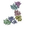
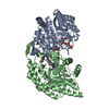
| Deposited unit | E: 2-methylcitrate dehydratase
D: 2-methylcitrate dehydratase
A: 2-methylcitrate dehydratase
B: 2-methylcitrate dehydratase
C: 2-methylcitrate dehydratase
F: 2-methylcitrate dehydratase
| Theoretical mass | Number of molelcules |
|---|
| Total (without water) | 322,550 | 6 |
|---|
| Polymers | 322,550 | 6 |
|---|
| Non-polymers | 0 | 0 |
|---|
| Water | 0 | 0 |
|---|
|
|---|
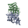
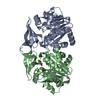
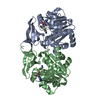
| 1 | E: 2-methylcitrate dehydratase
F: 2-methylcitrate dehydratase
| Theoretical mass | Number of molelcules |
|---|
| Total (without water) | 107,517 | 2 |
|---|
| Polymers | 107,517 | 2 |
|---|
| Non-polymers | 0 | 0 |
|---|
| Water | 0 | |
|---|
| Type | Name | Symmetry operation | Number |
|---|
| identity operation | 1_555 | x,y,z | 1 |
| Buried area | 4230 Å2 |
|---|
| ΔGint | -30 kcal/mol |
|---|
| Surface area | 24120 Å2 |
|---|
| Method | PISA |
|---|
|
|---|
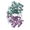
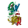
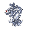
| 2 | D: 2-methylcitrate dehydratase
C: 2-methylcitrate dehydratase
| Theoretical mass | Number of molelcules |
|---|
| Total (without water) | 107,517 | 2 |
|---|
| Polymers | 107,517 | 2 |
|---|
| Non-polymers | 0 | 0 |
|---|
| Water | 0 | |
|---|
| Type | Name | Symmetry operation | Number |
|---|
| identity operation | 1_555 | x,y,z | 1 |
| Buried area | 4360 Å2 |
|---|
| ΔGint | -28 kcal/mol |
|---|
| Surface area | 24220 Å2 |
|---|
| Method | PISA |
|---|
|
|---|
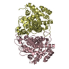
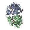
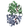
| 3 | A: 2-methylcitrate dehydratase
B: 2-methylcitrate dehydratase
| Theoretical mass | Number of molelcules |
|---|
| Total (without water) | 107,517 | 2 |
|---|
| Polymers | 107,517 | 2 |
|---|
| Non-polymers | 0 | 0 |
|---|
| Water | 0 | |
|---|
| Type | Name | Symmetry operation | Number |
|---|
| identity operation | 1_555 | x,y,z | 1 |
| Buried area | 4460 Å2 |
|---|
| ΔGint | -29 kcal/mol |
|---|
| Surface area | 28370 Å2 |
|---|
| Method | PISA |
|---|
|
|---|
| Unit cell | | Length a, b, c (Å) | 139.662, 139.662, 513.484 |
|---|
| Angle α, β, γ (deg.) | 90.00, 90.00, 120.00 |
|---|
| Int Tables number | 146 |
|---|
| Space group name H-M | H3 |
|---|
|
|---|
| Noncrystallographic symmetry (NCS) | NCS domain: | ID | Ens-ID | Details (eV) |
|---|
| 1 | 1 | E| 2 | 1 | D| 1 | 2 | E| 2 | 2 | A| 1 | 3 | E| 2 | 3 | B| 1 | 4 | E| 2 | 4 | C| 1 | 5 | E| 2 | 5 | F| 1 | 6 | D| 2 | 6 | A| 1 | 7 | D| 2 | 7 | B| 1 | 8 | D| 2 | 8 | C| 1 | 9 | D| 2 | 9 | F| 1 | 10 | A| 2 | 10 | B| 1 | 11 | A| 2 | 11 | C| 1 | 12 | A| 2 | 12 | F| 1 | 13 | B| 2 | 13 | C| 1 | 14 | B| 2 | 14 | F| 1 | 15 | C| 2 | 15 | F | | | | | | | | | | | | | | | | | | | | | | | | | | | | | |
NCS domain segments: Component-ID: _ / Beg auth comp-ID: ASP / Beg label comp-ID: ASP / Refine code: _ | Dom-ID | Ens-ID | End auth comp-ID | End label comp-ID | Auth asym-ID | Label asym-ID | Auth seq-ID | Label seq-ID |
|---|
| 1 | 1 | VALVALEA| 11 - 482 | 11 - 482 | | 2 | 1 | VALVALDB| 11 - 482 | 11 - 482 | | 1 | 2 | TYRTYREA| 11 - 481 | 11 - 481 | | 2 | 2 | PHEPHEAC| 11 - 358 | 11 - 358 | | 1 | 3 | VALVALEA| 11 - 482 | 11 - 482 | | 2 | 3 | VALVALBD| 11 - 482 | 11 - 482 | | 1 | 4 | VALVALEA| 11 - 482 | 11 - 482 | | 2 | 4 | VALVALCE| 11 - 482 | 11 - 482 | | 1 | 5 | VALVALEA| 11 - 482 | 11 - 482 | | 2 | 5 | VALVALFF| 11 - 482 | 11 - 482 | | 1 | 6 | TYRTYRDB| 11 - 481 | 11 - 481 | | 2 | 6 | PHEPHEAC| 11 - 358 | 11 - 358 | | 1 | 7 | VALVALDB| 11 - 482 | 11 - 482 | | 2 | 7 | VALVALBD| 11 - 482 | 11 - 482 | | 1 | 8 | VALVALDB| 11 - 482 | 11 - 482 | | 2 | 8 | VALVALCE| 11 - 482 | 11 - 482 | | 1 | 9 | VALVALDB| 11 - 482 | 11 - 482 | | 2 | 9 | VALVALFF| 11 - 482 | 11 - 482 | | 1 | 10 | PHEPHEA| C | | | | | | | | | | | | | | | | | | | | | | | | | | | | | | | | | | | | | | | | | | | | | | | | | | | | | | | | | | | | | | | | | | | | | | | | | | | |
|
|---|
 Movie
Movie Controller

 Yorodumi
 Open data
 Basic information
 Components
 Keywords
 Function and homology information
 Salmonella enterica (bacteria)
 X-RAY DIFFRACTION /
  Authors
 Citation
 Structure visualization
 Downloads & links
 Other downloads


 PDBj
PDBj

 Assembly