 Movie
Movie Controller

[English] 日本語
 Yorodumi
- PDB-1of8: double complex of the tyrosine sensitive DAHP Synthase from S. ce... -
+ Open data

- Basic information
| Entry | Database: PDB / ID: 1of8 | ||||||
|---|---|---|---|---|---|---|---|
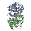
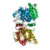
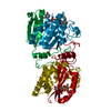
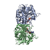
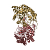
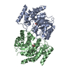
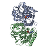
| Title | double complex of the tyrosine sensitive DAHP Synthase from S. cerevisiae with Co2+, PEP and the E4P analogoue G3P | ||||||
 Components Components | PHOSPHO-2-DEHYDRO-3-DEOXYHEPTONATE ALDOLASE, TYROSINE-INHIBITED | ||||||
 Keywords Keywords | LYASE / BETA-ALPHA-BARREL / SYNTHASE / ALDOLASE / SYNTHETASE | ||||||
| Function / homology |  Function and homology information Function and homology information3-deoxy-7-phosphoheptulonate synthase / 3-deoxy-7-phosphoheptulonate synthase activity / chorismate biosynthetic process / aromatic amino acid biosynthetic process / amino acid biosynthetic process / nucleus / cytoplasm Similarity search - Function | ||||||
| Biological species |  | ||||||
| Method |  X-RAY DIFFRACTION / SYNCHROTRON / MOLECULAR REPLACEMENT / Resolution: 1.5 Å X-RAY DIFFRACTION / SYNCHROTRON / MOLECULAR REPLACEMENT / Resolution: 1.5 Å | ||||||
 Authors Authors | Koenig, V. / Pfeil, A. / Heinrich, G. / Braus, G.H. / Schneider, T.R. | ||||||
 Citation Citation | Journal: J.Mol.Biol. / Year: 2004 Title: Substrate and Metal Complexes of 3-Deoxy-D-Arabino-Heptulosonate-7-Phosphate Synthase from Saccharomyces Cerevisiae Provide New Insights Into the Catalytic Mechanism Authors: Koenig, V. / Pfeil, A. / Braus, G.H. / Schneider, T.R. #1: Journal: Proc.Natl.Acad.Sci.USA / Year: 2003Title: Evolution of Feedback-Inhibited Beta /Alpha Barrel Isoenzymes by Gene Duplication and a Single Mutation. Authors: Hartmann, M. / Schneider, T.R. / Pfeil, A. / Heinrich, G. / Lipscomb, W.N. / Braus, G.H. | ||||||
| History |
| ||||||
| Remark 700 | SHEET DETERMINATION METHOD: DSSP THE SHEETS PRESENTED AS "AB" IN EACH CHAIN ON SHEET RECORDS BELOW ... SHEET DETERMINATION METHOD: DSSP THE SHEETS PRESENTED AS "AB" IN EACH CHAIN ON SHEET RECORDS BELOW IS ACTUALLY AN 8-STRANDED BARREL THIS IS REPRESENTED BY A 9-STRANDED SHEET IN WHICH THE FIRST AND LAST STRANDS ARE IDENTICAL. DETERMINATION METHOD: DSSP THE SHEETS PRESENTED AS "BA" IN EACH CHAIN ON SHEET RECORDS BELOW IS ACTUALLY AN 8-STRANDED BARREL THIS IS REPRESENTED BY A 9-STRANDED SHEET IN WHICH THE FIRST AND LAST STRANDS ARE IDENTICAL. |
- Structure visualization
| Structure viewer | Molecule: MolmilJmol/JSmol |
|---|
- Downloads & links
-Download
| PDBx/mmCIF format | 1of8.cif.gz | 297.6 KB | Display | PDBx/mmCIF format |
|---|---|---|---|---|
| PDB format | pdb1of8.ent.gz | 238.3 KB | Display | PDB format |
| PDBx/mmJSON format | 1of8.json.gz | Tree view | PDBx/mmJSON format | |
| Others |  Other downloads Other downloads |
-Validation report
| Arichive directory | https://data.pdbj.org/pub/pdb/validation_reports/of/1of8ftp://data.pdbj.org/pub/pdb/validation_reports/of/1of8 | HTTPS FTP |
|---|
-Related structure data
| Related structure data |  1oabC  1ofaC  1ofbC  1ofoC  1ofpC  1ofqC  1ofrC  1hfbS S: Starting model for refinement C: citing same article ( |
|---|---|
| Similar structure data |


-Links
 PDBj
PDBj

- Assembly
| Deposited unit | 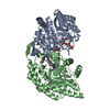
| ||||||||
|---|---|---|---|---|---|---|---|---|---|
| 1 |
| ||||||||
| Unit cell |
|
-Components
-Protein , 1 types, 2 molecules AB
| #1: Protein | Mass: 39797.055 Da / Num. of mol.: 2 Source method: isolated from a genetically manipulated source Source: (gene. exp.) Strain: RH1326 / Production host: |
|---|
-Non-polymers , 5 types, 798 molecules 


| #2: Chemical |  Mass: 58.933 Da / Num. of mol.: 2 / Source method: obtained synthetically / Formula: Co Mass: 58.933 Da / Num. of mol.: 2 / Source method: obtained synthetically / Formula: Co#3: Chemical |  Mass: 168.042 Da / Num. of mol.: 2 / Source method: obtained synthetically / Formula: C3H5O6P Mass: 168.042 Da / Num. of mol.: 2 / Source method: obtained synthetically / Formula: C3H5O6P#4: Chemical |  Mass: 172.074 Da / Num. of mol.: 2 / Source method: obtained synthetically / Formula: C3H9O6P Mass: 172.074 Da / Num. of mol.: 2 / Source method: obtained synthetically / Formula: C3H9O6P#5: Chemical |  Mass: 92.094 Da / Num. of mol.: 2 / Source method: obtained synthetically / Formula: C3H8O3 Mass: 92.094 Da / Num. of mol.: 2 / Source method: obtained synthetically / Formula: C3H8O3#6: Water | ChemComp-HOH / | Mass: 18.015 Da / Num. of mol.: 790 / Source method: isolated from a natural source / Formula: H2O |
|---|
-Experimental details
-Experiment
| Experiment | Method: X-RAY DIFFRACTION / Number of used crystals: 1 |
|---|
- Sample preparation
| Crystal | Density Matthews: 1.95 Å3/Da / Density % sol: 36.77 % |
|---|---|
| Crystal grow | pH: 8.5 / Details: pH 8.50 |
-Data collection
| Diffraction | Mean temperature: 100 K |
|---|---|
| Diffraction source | Source: SYNCHROTRON / Site: EMBL/DESY, HAMBURG  / Beamline: X11 / Wavelength: 0.811 / Beamline: X11 / Wavelength: 0.811 |
| Detector | Type: MARRESEARCH / Detector: CCD / Date: Jul 7, 2002 / Details: MIRRORS |
| Radiation | Monochromator: GRAPHITE / Protocol: SINGLE WAVELENGTH / Monochromatic (M) / Laue (L): M / Scattering type: x-ray |
| Radiation wavelength | Wavelength: 0.811 Å / Relative weight: 1 |
| Reflection | Resolution: 1.5→27.3 Å / Num. obs: 234883 / % possible obs: 96.6 % / Observed criterion σ(I): 1.5 / Redundancy: 2.4 % / Biso Wilson estimate: 16.1 Å2 / Rmerge(I) obs: 0.057 / Net I/σ(I): 9.75 |
| Reflection shell | Resolution: 1.5→1.55 Å / Redundancy: 1.98 % / Rmerge(I) obs: 0.32 / Mean I/σ(I) obs: 2.82 / % possible all: 96.3 |
- Processing
| Software |
| ||||||||||||||||||||
|---|---|---|---|---|---|---|---|---|---|---|---|---|---|---|---|---|---|---|---|---|---|
| Refinement | Method to determine structure: MOLECULAR REPLACEMENT Starting model: MOLECULE A OF PDB ENTRY 1HFB Resolution: 1.5→27 Å / SU B: 1.119 / SU ML: 0.042 / Cross valid method: THROUGHOUT / ESU R: 0.078 / ESU R Free: 0.066
| ||||||||||||||||||||
| Displacement parameters | Biso mean: 14.747 Å2
| ||||||||||||||||||||
| Refinement step | Cycle: LAST / Resolution: 1.5→27 Å
|
