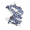
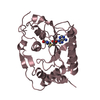
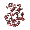
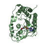
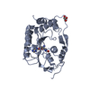
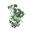
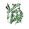
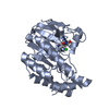
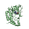
Entry Database : PDB / ID : 5mrkTitle Structural basis of Zika virus methyltransferase inhibition by sinefungin methyltransferase Keywords / / / / Function / homology Function Domain/homology Component
/ / / / / / / / / / / / / / / / / / / / / / / / / / / / / / / / / / / / / / / / / / / / / / / / / / / / / / / / / / / / / / / / / / / / / / / / / / / / / / / / / / / / / / / / / / / / / / / / / / / / / / / / / / / / / / / / / / / / / / / / / / / / / Biological species Method / / Resolution : 1.9 Å Authors Hercik, K. / Boura, E. Journal : Arch. Virol. / Year : 2017Title : Structural basis of Zika virus methyltransferase inhibition by sinefungin.Authors : Hercik, K. / Brynda, J. / Nencka, R. / Boura, E. History Deposition Dec 23, 2016 Deposition site / Processing site Revision 1.0 Jan 25, 2017 Provider / Type Revision 1.1 May 17, 2017 Group Revision 1.2 Jun 28, 2017 Group / Category Item / _citation.page_first / _citation.page_lastRevision 1.3 Jan 17, 2024 Group Advisory / Data collection ... Advisory / Data collection / Database references / Refinement description Category chem_comp_atom / chem_comp_bond ... chem_comp_atom / chem_comp_bond / database_2 / pdbx_initial_refinement_model / pdbx_unobs_or_zero_occ_atoms Item / _database_2.pdbx_database_accessionRevision 1.4 Oct 23, 2024 Group / Category / pdbx_modification_feature
Show all Show less
 Movie
Movie Controller

 Yorodumi
 Open data
 Basic information
 Components
 Keywords
 Function and homology information
 Zika virus
 X-RAY DIFFRACTION /
  Authors
 Citation
 Structure visualization
 Downloads & links
 Other downloads
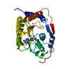
 PDBj
PDBj


 Assembly
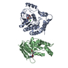


 Mass: 381.387 Da / Num. of mol.: 2 / Source method: obtained synthetically / Formula: C15H23N7O5
 Mass: 35.453 Da / Num. of mol.: 2 / Source method: isolated from a natural source / Formula: Cl
 Mass: 18.015 Da / Num. of mol.: 327 / Source method: isolated from a natural source / Formula: H2O
 Sample preparation
 Processing