 Movie
Movie Controller

[English] 日本語
 Yorodumi
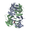
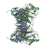
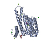
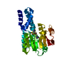
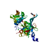
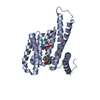
- PDB-5mqh: Structure of the Phosphatase Domain of the Cell Fate Determinant ... -
+ Open data

- Basic information
| Entry | Database: PDB / ID: 5mqh | ||||||
|---|---|---|---|---|---|---|---|
| Title | Structure of the Phosphatase Domain of the Cell Fate Determinant SpoIIE from Bacillus subtilis in a crystal form without domain swapping | ||||||
 Components Components | Serine phosphatase | ||||||
 Keywords Keywords | TRANSFERASE / Sporulation / Phosphatase / PP2C / Manganese | ||||||
| Function / homology |  Function and homology information Function and homology informationendospore-forming forespore / sporulation resulting in formation of a cellular spore / protein-serine/threonine phosphatase / protein serine/threonine phosphatase activity / phosphatase activity / membrane / plasma membrane Similarity search - Function | ||||||
| Biological species |  | ||||||
| Method |  X-RAY DIFFRACTION / SYNCHROTRON / MOLECULAR REPLACEMENT / Resolution: 2.45 Å X-RAY DIFFRACTION / SYNCHROTRON / MOLECULAR REPLACEMENT / Resolution: 2.45 Å | ||||||
 Authors Authors | Levdikov, V.M. / Wilkinson, A.J. / Blagova, E.V. | ||||||
| Funding support |  United Kingdom, 1items United Kingdom, 1items
| ||||||
 Citation Citation | Journal: Elife / Year: 2017 Title: A widespread family of serine/threonine protein phosphatases shares a common regulatory switch with proteasomal proteases. Authors: Bradshaw, N. / Levdikov, V.M. / Zimanyi, C.M. / Gaudet, R. / Wilkinson, A.J. / Losick, R. #1: Journal: J. Mol. Biol. / Year: 2012Title: Structure of the phosphatase domain of the cell fate determinant SpoIIE from Bacillus subtilis. Authors: Levdikov, V.M. / Blagova, E.V. / Rawlings, A.E. / Jameson, K. / Tunaley, J. / Hart, D.J. / Barak, I. / Wilkinson, A.J. #2: Journal: Elife / Year: 2015 Title: Asymmetric division triggers cell-specific gene expression through coupled capture and stabilization of a phosphatase. Authors: Bradshaw, N. / Losick, R. | ||||||
| History |
|
- Structure visualization
| Structure viewer | Molecule: MolmilJmol/JSmol |
|---|
- Downloads & links
-Download
| PDBx/mmCIF format | 5mqh.cif.gz | 59.8 KB | Display | PDBx/mmCIF format |
|---|---|---|---|---|
| PDB format | pdb5mqh.ent.gz | 42.4 KB | Display | PDB format |
| PDBx/mmJSON format | 5mqh.json.gz | Tree view | PDBx/mmJSON format | |
| Others |  Other downloads Other downloads |
-Validation report
| Arichive directory | https://data.pdbj.org/pub/pdb/validation_reports/mq/5mqhftp://data.pdbj.org/pub/pdb/validation_reports/mq/5mqh | HTTPS FTP |
|---|
-Related structure data
| Related structure data | 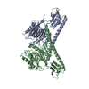 5ucgC  3t9lS S: Starting model for refinement C: citing same article ( |
|---|---|
| Similar structure data |


-Links
 PDBj
PDBj- Assembly
| Deposited unit | 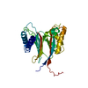
| ||||||||
|---|---|---|---|---|---|---|---|---|---|
| 1 |
| ||||||||
| Unit cell |
| ||||||||
| Components on special symmetry positions |
|

-Components
| #1: Protein | Mass: 27417.348 Da / Num. of mol.: 1 / Mutation: A624I Source method: isolated from a genetically manipulated source Details: The mutation Ala624 to Ile was introduced to reduce successfully the extent of domain swapping. Source: (gene. exp.) |
|---|---|
| #2: Water | ChemComp-HOH /  Mass: 18.015 Da / Num. of mol.: 33 / Source method: isolated from a natural source / Formula: H2O Mass: 18.015 Da / Num. of mol.: 33 / Source method: isolated from a natural source / Formula: H2O |
-Experimental details
-Experiment
| Experiment | Method: X-RAY DIFFRACTION / Number of used crystals: 1 |
|---|
- Sample preparation
| Crystal | Density Matthews: 2.57 Å3/Da / Density % sol: 52.13 % |
|---|---|
| Crystal grow | Temperature: 291 K / Method: vapor diffusion, hanging drop / pH: 4.6 / Details: 2M Sodium formate, 0.1M Sodium acetate |
-Data collection
| Diffraction | Mean temperature: 100 K |
|---|---|
| Diffraction source | Source: SYNCHROTRON / Site: Diamond / Beamline: I02 / Wavelength: 0.9795 Å |
| Detector | Type: DECTRIS PILATUS3 S 6M / Detector: PIXEL / Date: Dec 12, 2012 |
| Radiation | Protocol: SINGLE WAVELENGTH / Monochromatic (M) / Laue (L): M / Scattering type: x-ray |
| Radiation wavelength | Wavelength: 0.9795 Å / Relative weight: 1 |
| Reflection | Resolution: 2.44→61.34 Å / Num. obs: 10961 / % possible obs: 99.7 % / Redundancy: 6.3 % / CC1/2: 0.999 / Rsym value: 0.057 / Net I/σ(I): 20.1 |
| Reflection shell | Resolution: 2.44→2.48 Å / Redundancy: 6.2 % / Mean I/σ(I) obs: 2.8 / CC1/2: 0.874 / % possible all: 99.8 |
- Processing
| Software |
| ||||||||||||||||||||||||||||||||||||||||||||||||||||||||||||||||||||||||||||||||||||||||||||||||||||||||||||||||||||||||||||||||||||||||||||||||||||||||||||||||||||||||||||||||||||||
|---|---|---|---|---|---|---|---|---|---|---|---|---|---|---|---|---|---|---|---|---|---|---|---|---|---|---|---|---|---|---|---|---|---|---|---|---|---|---|---|---|---|---|---|---|---|---|---|---|---|---|---|---|---|---|---|---|---|---|---|---|---|---|---|---|---|---|---|---|---|---|---|---|---|---|---|---|---|---|---|---|---|---|---|---|---|---|---|---|---|---|---|---|---|---|---|---|---|---|---|---|---|---|---|---|---|---|---|---|---|---|---|---|---|---|---|---|---|---|---|---|---|---|---|---|---|---|---|---|---|---|---|---|---|---|---|---|---|---|---|---|---|---|---|---|---|---|---|---|---|---|---|---|---|---|---|---|---|---|---|---|---|---|---|---|---|---|---|---|---|---|---|---|---|---|---|---|---|---|---|---|---|---|---|
| Refinement | Method to determine structure: MOLECULAR REPLACEMENT Starting model: 3T9L Resolution: 2.45→48.99 Å / Cor.coef. Fo:Fc: 0.951 / Cor.coef. Fo:Fc free: 0.94 / SU B: 13.871 / SU ML: 0.296 / Cross valid method: THROUGHOUT / ESU R: 0.402 / ESU R Free: 0.29 / Details: HYDROGENS HAVE BEEN ADDED IN THE RIDING POSITIONS
| ||||||||||||||||||||||||||||||||||||||||||||||||||||||||||||||||||||||||||||||||||||||||||||||||||||||||||||||||||||||||||||||||||||||||||||||||||||||||||||||||||||||||||||||||||||||
| Solvent computation | Ion probe radii: 0.8 Å / Shrinkage radii: 0.8 Å / VDW probe radii: 1.2 Å | ||||||||||||||||||||||||||||||||||||||||||||||||||||||||||||||||||||||||||||||||||||||||||||||||||||||||||||||||||||||||||||||||||||||||||||||||||||||||||||||||||||||||||||||||||||||
| Displacement parameters | Biso mean: 66.958 Å2
| ||||||||||||||||||||||||||||||||||||||||||||||||||||||||||||||||||||||||||||||||||||||||||||||||||||||||||||||||||||||||||||||||||||||||||||||||||||||||||||||||||||||||||||||||||||||
| Refinement step | Cycle: 1 / Resolution: 2.45→48.99 Å
| ||||||||||||||||||||||||||||||||||||||||||||||||||||||||||||||||||||||||||||||||||||||||||||||||||||||||||||||||||||||||||||||||||||||||||||||||||||||||||||||||||||||||||||||||||||||
| Refine LS restraints |
|