 Movie
Movie Controller


+ Open data

- Basic information
| Entry | Database: PDB / ID: 5mqc | ||||||
|---|---|---|---|---|---|---|---|
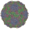
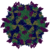
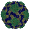
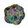
| Title | Structure of black queen cell virus | ||||||
 Components Components |
| ||||||
 Keywords Keywords | VIRUS / Apis mellifera / honey bee / honeybee / Picornavirales / Dicistroviridae / Cripavirus / virion / capsid | ||||||
| Function / homology |  Function and homology information Function and homology information | ||||||
| Biological species |  Black queen cell virus Black queen cell virus | ||||||
| Method |  X-RAY DIFFRACTION / SYNCHROTRON / MOLECULAR REPLACEMENT / Resolution: 3.4 Å X-RAY DIFFRACTION / SYNCHROTRON / MOLECULAR REPLACEMENT / Resolution: 3.4 Å | ||||||
 Authors Authors | Spurny, R. / Kiem, H.H.T. / Plevka, P. | ||||||
| Funding support |  Czech Republic, 1items Czech Republic, 1items
| ||||||
 Citation Citation | Journal: J. Virol. / Year: 2017 Title: Virion Structure of Black Queen Cell Virus, a Common Honeybee Pathogen. Authors: Spurny, R. / Pridal, A. / Palkova, L. / Kiem, H.K. / de Miranda, J.R. / Plevka, P. | ||||||
| History |
|
- Structure visualization
| Structure viewer | Molecule: MolmilJmol/JSmol |
|---|
- Downloads & links
-Download
| PDBx/mmCIF format | 5mqc.cif.gz | 161.2 KB | Display | PDBx/mmCIF format |
|---|---|---|---|---|
| PDB format | pdb5mqc.ent.gz | 126.8 KB | Display | PDB format |
| PDBx/mmJSON format | 5mqc.json.gz | Tree view | PDBx/mmJSON format | |
| Others |  Other downloads Other downloads |
-Validation report
| Arichive directory | https://data.pdbj.org/pub/pdb/validation_reports/mq/5mqcftp://data.pdbj.org/pub/pdb/validation_reports/mq/5mqc | HTTPS FTP |
|---|
-Related structure data
| Related structure data |  1b35S S: Starting model for refinement |
|---|---|
| Similar structure data |
-Links
 PDBj
PDBj
- Assembly
| Deposited unit | 
| ||||||||
|---|---|---|---|---|---|---|---|---|---|
| 1 | x 60
| ||||||||
| 2 |
| ||||||||
| 3 | x 5
| ||||||||
| 4 | x 6
| ||||||||
| 5 | 
| ||||||||
| Unit cell |
| ||||||||
| Symmetry | Point symmetry: (Schoenflies symbol: I (icosahedral)) |
-Components
| #1: Protein | Mass: 31222.748 Da / Num. of mol.: 1 / Source method: isolated from a natural source / Source: (natural) Black queen cell virus / References: UniProt: Q9J7C2 |
|---|---|
| #2: Protein | Mass: 30108.285 Da / Num. of mol.: 1 / Source method: isolated from a natural source / Source: (natural) Black queen cell virus / References: UniProt: Q9J7C2 |
| #3: Protein | Mass: 26297.910 Da / Num. of mol.: 1 / Source method: isolated from a natural source / Source: (natural) Black queen cell virus / References: UniProt: Q9J7C2 |
-Experimental details
-Experiment
| Experiment | Method: X-RAY DIFFRACTION / Number of used crystals: 1 |
|---|
- Sample preparation
| Crystal grow | Temperature: 295 K / Method: vapor diffusion, hanging drop |
|---|
-Data collection
| Diffraction | Mean temperature: 80 K |
|---|---|
| Diffraction source | Source: SYNCHROTRON / Site: SOLEIL  / Beamline: PROXIMA 1 / Wavelength: 1 Å / Beamline: PROXIMA 1 / Wavelength: 1 Å |
| Detector | Type: DECTRIS PILATUS 6M / Detector: PIXEL / Date: Dec 2, 2015 |
| Radiation | Protocol: SINGLE WAVELENGTH / Monochromatic (M) / Laue (L): M / Scattering type: x-ray |
| Radiation wavelength | Wavelength: 1 Å / Relative weight: 1 |
| Reflection | Resolution: 3.4→39.81 Å / Num. obs: 193433 / % possible obs: 68.1 % / Redundancy: 1.9 % / Rmerge(I) obs: 0.208 / Net I/σ(I): 3.7 |
- Processing
| Software |
| ||||||||||||
|---|---|---|---|---|---|---|---|---|---|---|---|---|---|
| Refinement | Method to determine structure: MOLECULAR REPLACEMENT Starting model: 1b35 Resolution: 3.4→39.81 Å / Cross valid method: NONE
| ||||||||||||
| Refinement step | Cycle: LAST / Resolution: 3.4→39.81 Å
|
