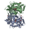
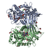
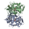
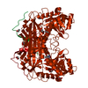
[English] 日本語
Yorodumi
- PDB-5mpr: Single Amino Acid Variant of Human Mitochondrial Branched Chain A... -

+
Open data


ID or keywords:

Loading...

-
Basic information

Entry
Database: PDB / ID: 5mpr
TitleSingle Amino Acid Variant of Human Mitochondrial Branched Chain Amino Acid Aminotransferase 2
ComponentsBranched-chain-amino-acid aminotransferase, mitochondrial
KeywordsTRANSFERASE / aminotransferase transaminase PLP mutation
Function / homology
Function and homology information


regulation of hormone levels / isoleucine catabolic process / L-leucine:2-oxoglutarate aminotransferase activity / branched-chain-amino-acid transaminase / L-leucine transaminase activity / L-valine transaminase activity / L-isoleucine transaminase activity / branched-chain amino acid biosynthetic process / branched-chain-amino-acid transaminase activity / Branched-chain amino acid catabolism ...regulation of hormone levels / isoleucine catabolic process / L-leucine:2-oxoglutarate aminotransferase activity / branched-chain-amino-acid transaminase / L-leucine transaminase activity / L-valine transaminase activity / L-isoleucine transaminase activity / branched-chain amino acid biosynthetic process / branched-chain-amino-acid transaminase activity / Branched-chain amino acid catabolism / L-leucine biosynthetic process / valine biosynthetic process / amino acid biosynthetic process / cellular response to leukemia inhibitory factor / lipid metabolic process / mitochondrial matrix / mitochondrion / nucleoplasm
Similarity search - Function
Branched-chain amino acid aminotransferase II / Branched-chain aminotransferase / Aminotransferase, class IV, conserved site / Aminotransferases class-IV signature. / Aminotransferase class 4, branched-chain amino acid transferase, N-terminal domain / D-amino Acid Aminotransferase; Chain A, domain 2 / D-amino Acid Aminotransferase, subunit A, domain 2 / Aminotransferase class IV / Aminotransferase-like, PLP-dependent enzymes / Branched-chain-amino-acid aminotransferase-like, N-terminal ...Branched-chain amino acid aminotransferase II / Branched-chain aminotransferase / Aminotransferase, class IV, conserved site / Aminotransferases class-IV signature. / Aminotransferase class 4, branched-chain amino acid transferase, N-terminal domain / D-amino Acid Aminotransferase; Chain A, domain 2 / D-amino Acid Aminotransferase, subunit A, domain 2 / Aminotransferase class IV / Aminotransferase-like, PLP-dependent enzymes / Branched-chain-amino-acid aminotransferase-like, N-terminal / Branched-chain-amino-acid aminotransferase-like, C-terminal / Amino-transferase class IV / D-amino Acid Aminotransferase; Chain A, domain 1 / Alpha-Beta Barrel / 2-Layer Sandwich / Alpha Beta
Similarity search - Domain/homology
PYRIDOXAL-5'-PHOSPHATE / Branched-chain-amino-acid aminotransferase, mitochondrial
Similarity search - Component
Biological speciesHomo sapiens (human)
MethodX-RAY DIFFRACTION / SYNCHROTRON / Resolution: 1.6 Å
AuthorsHakansson, M. / Walse, B. / Nilsson, C. / Anderson, L.C.
Funding support Sweden, 1items
OrganizationGrant numberCountry
Sweden
CitationJournal: J. Am. Soc. Mass Spectrom. / Year: 2017
Title: Intact Protein Analysis at 21 Tesla and X-Ray Crystallography Define Structural Differences in Single Amino Acid Variants of Human Mitochondrial Branched-Chain Amino Acid Aminotransferase 2 (BCAT2).
Authors: Anderson, L.C. / Hakansson, M. / Walse, B. / Nilsson, C.L.
History
DepositionDec 18, 2016Deposition site: PDBE / Processing site: PDBE
Revision 1.0Jul 19, 2017Provider: repository / Type: Initial release
Revision 1.1Dec 13, 2017Group: Database references / Category: citation
Item: _citation.journal_volume / _citation.page_first / _citation.page_last
Revision 1.2Oct 16, 2019Group: Data collection / Category: reflns_shell

-
Structure visualization

Structure viewerMolecule:
MolmilJmol/JSmol

Downloads & links

-
Assembly

Deposited unit
A: Branched-chain-amino-acid aminotransferase, mitochondrial
hetero molecules


Theoretical massNumber of molelcules
Total (without water)41,9837
Polymers41,4251
Non-polymers5576
Water4,252236
1
A: Branched-chain-amino-acid aminotransferase, mitochondrial
hetero molecules

A: Branched-chain-amino-acid aminotransferase, mitochondrial
hetero molecules


Theoretical massNumber of molelcules
Total (without water)83,96514
Polymers82,8502
Non-polymers1,11512
Water362
TypeNameSymmetry operationNumber
identity operation1_555x,y,z1
crystal symmetry operation5_677x-y+1,-y+2,-z+7/31
Buried area8080 Å2
ΔGint6 kcal/mol
Surface area27770 Å2
MethodPISA
Unit cell
Length a, b, c (Å)84.032, 84.032, 105.446
Angle α, β, γ (deg.)90.00, 90.00, 120.00
Int Tables number154
Space group name H-MP3221

-
Components

#1: Protein Branched-chain-amino-acid aminotransferase, mitochondrial / BCAT(m) / Placental protein 18 / PP18


Mass: 41425.062 Da / Num. of mol.: 1 / Mutation: T159R
Source method: isolated from a genetically manipulated source
Source: (gene. exp.) Homo sapiens (human) / Gene: BCAT2, BCATM, BCT2, ECA40 / Production host: Escherichia coli (E. coli)
References: UniProt: O15382, branched-chain-amino-acid transaminase
#2: Chemical ChemComp-PLP / PYRIDOXAL-5'-PHOSPHATE / VITAMIN B6 Phosphate


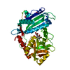
Mass: 247.142 Da / Num. of mol.: 1 / Source method: obtained synthetically / Formula: C8H10NO6P
#3: Chemical
ChemComp-EDO / 1,2-ETHANEDIOL / ETHYLENE GLYCOL


Mass: 62.068 Da / Num. of mol.: 5 / Source method: obtained synthetically / Formula: C2H6O2
#4: Water ChemComp-HOH / water


Mass: 18.015 Da / Num. of mol.: 236 / Source method: isolated from a natural source / Formula: H2O

-
Experimental details

-
Experiment

ExperimentMethod: X-RAY DIFFRACTION / Number of used crystals: 1

-
Sample preparation

CrystalDensity Matthews: 2.59 Å3/Da / Density % sol: 52.59 %
Crystal growTemperature: 293 K / Method: evaporation / pH: 6.5
Details: 0.2 M Sodiumfluoride, 0.1 M Bis Tris propane pH 6.5, 20% w/v Polyethylene glycol 3350

-
Data collection

DiffractionMean temperature: 100 K
Diffraction sourceSource: SYNCHROTRON / Site: Diamond / Beamline: I03 / Wavelength: 0.97625 Å
DetectorType: DECTRIS PILATUS3 S 6M / Detector: PIXEL / Date: Apr 8, 2016
RadiationProtocol: SINGLE WAVELENGTH / Monochromatic (M) / Laue (L): M / Scattering type: x-ray
Radiation wavelengthWavelength: 0.97625 Å / Relative weight: 1
ReflectionResolution: 1.6→28 Å / Num. obs: 57390 / % possible obs: 99.7 % / Redundancy: 9.7 % / Net I/σ(I): 11.8

-
Processing

Software
NameVersionClassification
REFMAC5.7.0032refinement
XDSdata reduction
Aimlessdata scaling
PHASERphasing
RefinementResolution: 1.6→27.52 Å / Cor.coef. Fo:Fc: 0.983 / Cor.coef. Fo:Fc free: 0.971 / SU B: 3.771 / SU ML: 0.056 / Cross valid method: THROUGHOUT / ESU R: 0.075 / ESU R Free: 0.072 / Details: HYDROGENS HAVE BEEN ADDED IN THE RIDING POSITIONS
RfactorNum. reflection% reflectionSelection details
Rfree0.17939 2912 5.1 %RANDOM
Rwork0.1279 ---
obs0.13052 54393 99.66 %-
Solvent computationIon probe radii: 0.8 Å / Shrinkage radii: 0.8 Å / VDW probe radii: 1.2 Å
Displacement parametersBiso mean: 28.859 Å2
Baniso -1Baniso -2Baniso -3
1-0.2 Å20.2 Å20 Å2
2--0.2 Å20 Å2
3----0.65 Å2
Refinement stepCycle: 1 / Resolution: 1.6→27.52 Å
ProteinNucleic acidLigandSolventTotal
Num. atoms2901 0 35 236 3172
Refine LS restraints
Refine-IDTypeDev idealDev ideal targetNumber
X-RAY DIFFRACTIONr_bond_refined_d0.0180.0193123
X-RAY DIFFRACTIONr_bond_other_d0.0010.023036
X-RAY DIFFRACTIONr_angle_refined_deg1.8161.9754250
X-RAY DIFFRACTIONr_angle_other_deg0.87536984
X-RAY DIFFRACTIONr_dihedral_angle_1_deg5.9265387
X-RAY DIFFRACTIONr_dihedral_angle_2_deg31.8223.066137
X-RAY DIFFRACTIONr_dihedral_angle_3_deg13.27515533
X-RAY DIFFRACTIONr_dihedral_angle_4_deg16.1561525
X-RAY DIFFRACTIONr_chiral_restr0.1160.2458
X-RAY DIFFRACTIONr_gen_planes_refined0.010.0213516
X-RAY DIFFRACTIONr_gen_planes_other0.0030.02736
X-RAY DIFFRACTIONr_nbd_refined
X-RAY DIFFRACTIONr_nbd_other
X-RAY DIFFRACTIONr_nbtor_refined
X-RAY DIFFRACTIONr_nbtor_other
X-RAY DIFFRACTIONr_xyhbond_nbd_refined
X-RAY DIFFRACTIONr_xyhbond_nbd_other
X-RAY DIFFRACTIONr_metal_ion_refined
X-RAY DIFFRACTIONr_metal_ion_other
X-RAY DIFFRACTIONr_symmetry_vdw_refined
X-RAY DIFFRACTIONr_symmetry_vdw_other
X-RAY DIFFRACTIONr_symmetry_hbond_refined
X-RAY DIFFRACTIONr_symmetry_hbond_other
X-RAY DIFFRACTIONr_symmetry_metal_ion_refined
X-RAY DIFFRACTIONr_symmetry_metal_ion_other
X-RAY DIFFRACTIONr_mcbond_it4.5132.4111503
X-RAY DIFFRACTIONr_mcbond_other4.4972.411502
X-RAY DIFFRACTIONr_mcangle_it5.2983.6311890
X-RAY DIFFRACTIONr_mcangle_other5.3053.6321891
X-RAY DIFFRACTIONr_scbond_it6.8173.0341615
X-RAY DIFFRACTIONr_scbond_other6.8163.0351615
X-RAY DIFFRACTIONr_scangle_it
X-RAY DIFFRACTIONr_scangle_other8.0254.312350
X-RAY DIFFRACTIONr_long_range_B_refined7.22821.1483544
X-RAY DIFFRACTIONr_long_range_B_other7.22821.1523545
X-RAY DIFFRACTIONr_rigid_bond_restr6.29836152
X-RAY DIFFRACTIONr_sphericity_free29.486579
X-RAY DIFFRACTIONr_sphericity_bonded17.96556229
LS refinement shellResolution: 1.598→1.639 Å / Total num. of bins used: 20
RfactorNum. reflection% reflection
Rfree0.346 196 -
Rwork0.248 3806 -
obs--95.7 %

+
About Yorodumi

-
News

-
Feb 9, 2022. New format data for meta-information of EMDB entries

New format data for meta-information of EMDB entries

  • Version 3 of the EMDB header file is now the official format.
  • The previous official version 1.9 will be removed from the archive.

Related info.:EMDB header

External links:wwPDB to switch to version 3 of the EMDB data model

-
Aug 12, 2020. Covid-19 info

Covid-19 info

URL: https://pdbjlvh1.pdbj.org/emnavi/covid19.php

New page: Covid-19 featured information page in EM Navigator.

Related info.:Covid-19 info / Mar 5, 2020. Novel coronavirus structure data

+
Mar 5, 2020. Novel coronavirus structure data

Novel coronavirus structure data

Related info.:Yorodumi Speices / Aug 12, 2020. Covid-19 info

External links:COVID-19 featured content - PDBj / Molecule of the Month (242):Coronavirus Proteases

+
Jan 31, 2019. EMDB accession codes are about to change! (news from PDBe EMDB page)

EMDB accession codes are about to change! (news from PDBe EMDB page)

  • The allocation of 4 digits for EMDB accession codes will soon come to an end. Whilst these codes will remain in use, new EMDB accession codes will include an additional digit and will expand incrementally as the available range of codes is exhausted. The current 4-digit format prefixed with “EMD-” (i.e. EMD-XXXX) will advance to a 5-digit format (i.e. EMD-XXXXX), and so on. It is currently estimated that the 4-digit codes will be depleted around Spring 2019, at which point the 5-digit format will come into force.
  • The EM Navigator/Yorodumi systems omit the EMD- prefix.

Related info.:Q: What is EMD? / ID/Accession-code notation in Yorodumi/EM Navigator

External links:EMDB Accession Codes are Changing Soon! / Contact to PDBj

+
Jul 12, 2017. Major update of PDB

Major update of PDB

  • wwPDB released updated PDB data conforming to the new PDBx/mmCIF dictionary.
  • This is a major update changing the version number from 4 to 5, and with Remediation, in which all the entries are updated.
  • In this update, many items about electron microscopy experimental information are reorganized (e.g. em_software).
  • Now, EM Navigator and Yorodumi are based on the updated data.

External links:wwPDB Remediation / Enriched Model Files Conforming to OneDep Data Standards Now Available in the PDB FTP Archive

-
Yorodumi

Thousand views of thousand structures

  • Yorodumi is a browser for structure data from EMDB, PDB, SASBDB, etc.
  • This page is also the successor to EM Navigator detail page, and also detail information page/front-end page for Omokage search.
  • The word "yorodu" (or yorozu) is an old Japanese word meaning "ten thousand". "mi" (miru) is to see.

Related info.:EMDB / PDB / SASBDB / Comparison of 3 databanks / Yorodumi Search / Aug 31, 2016. New EM Navigator & Yorodumi / Yorodumi Papers / Jmol/JSmol / Function and homology information / Changes in new EM Navigator and Yorodumi

Read more