 Movie
Movie Controller

[English] 日本語
 Yorodumi
- PDB-5mm1: Dolichyl phosphate mannose synthase in complex with GDP and dolic... -
+ Open data

- Basic information
| Entry | Database: PDB / ID: 5mm1 | ||||||||||||
|---|---|---|---|---|---|---|---|---|---|---|---|---|---|
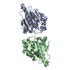
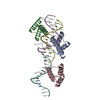
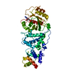
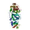
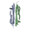
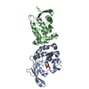
| Title | Dolichyl phosphate mannose synthase in complex with GDP and dolichyl phosphate mannose | ||||||||||||
 Components Components | Dolichol monophosphate mannose synthase | ||||||||||||
 Keywords Keywords | MEMBRANE PROTEIN / Dolichol phosphate mannose synthase / enzyme / integral membrane protein / protein glycosylation / product complex / GDP / dolichyl phosphate mannose | ||||||||||||
| Function / homology |  Function and homology information Function and homology informationdolichyl-phosphate beta-D-mannosyltransferase / dolichyl-phosphate beta-D-mannosyltransferase activity / dolichol phosphate mannose biosynthetic process / GPI anchor biosynthetic process / dolichol-linked oligosaccharide biosynthetic process / protein O-linked glycosylation via mannose / polysaccharide biosynthetic process / manganese ion binding / magnesium ion binding / metal ion binding / plasma membrane Similarity search - Function | ||||||||||||
| Biological species |   Pyrococcus furiosus (archaea) Pyrococcus furiosus (archaea) | ||||||||||||
| Method |  X-RAY DIFFRACTION / SYNCHROTRON / MOLECULAR REPLACEMENT / Resolution: 2.6 Å X-RAY DIFFRACTION / SYNCHROTRON / MOLECULAR REPLACEMENT / Resolution: 2.6 Å | ||||||||||||
 Authors Authors | Gandini, R. / Reichenbach, T. / Tan, T.C. / Divne, C. | ||||||||||||
| Funding support |  Sweden, 3items Sweden, 3items
| ||||||||||||
 Citation Citation | Journal: Nat Commun / Year: 2017 Title: Structural basis for dolichylphosphate mannose biosynthesis. Authors: Gandini, R. / Reichenbach, T. / Tan, T.C. / Divne, C. | ||||||||||||
| History |
|
- Structure visualization
| Structure viewer | Molecule: MolmilJmol/JSmol |
|---|
- Downloads & links
-Download
| PDBx/mmCIF format | 5mm1.cif.gz | 88.3 KB | Display | PDBx/mmCIF format |
|---|---|---|---|---|
| PDB format | pdb5mm1.ent.gz | 64.3 KB | Display | PDB format |
| PDBx/mmJSON format | 5mm1.json.gz | Tree view | PDBx/mmJSON format | |
| Others |  Other downloads Other downloads |
-Validation report
| Arichive directory | https://data.pdbj.org/pub/pdb/validation_reports/mm/5mm1ftp://data.pdbj.org/pub/pdb/validation_reports/mm/5mm1 | HTTPS FTP |
|---|
-Related structure data
| Related structure data | 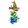 5mlzSC 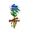 5mm0C S: Starting model for refinement C: citing same article ( |
|---|---|
| Similar structure data |


-Links
 PDBj
PDBj

- Assembly
| Deposited unit | 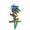
| ||||||||
|---|---|---|---|---|---|---|---|---|---|
| 1 |
| ||||||||
| Unit cell |
|
-Components
| #1: Protein | Mass: 42684.809 Da / Num. of mol.: 1 Source method: isolated from a genetically manipulated source Details: N-terminal hexa-histdine tag and TEV protease cleavage site precedes the protein sequences Source: (gene. exp.) Pyrococcus furiosus (strain ATCC 43587 / DSM 3638 / JCM 8422 / Vc1) (archaea)Gene: PF0058 / Plasmid: pNIC28-Bsa4 / Production host:  |
|---|---|
| #2: Chemical | ChemComp-GDP /   Type: RNA linking / Mass: 443.201 Da / Num. of mol.: 1 / Source method: obtained synthetically / Formula: C10H15N5O11P2 / Comment: GDP, energy-carrying molecule*YM Type: RNA linking / Mass: 443.201 Da / Num. of mol.: 1 / Source method: obtained synthetically / Formula: C10H15N5O11P2 / Comment: GDP, energy-carrying molecule*YM |
| #3: Chemical | ChemComp-MJC /   Mass: 1011.439 Da / Num. of mol.: 1 / Source method: obtained synthetically / Formula: C61H103O9P Mass: 1011.439 Da / Num. of mol.: 1 / Source method: obtained synthetically / Formula: C61H103O9P |
-Experimental details
-Experiment
| Experiment | Method: X-RAY DIFFRACTION / Number of used crystals: 1 |
|---|
- Sample preparation
| Crystal | Density Matthews: 3.69 Å3/Da / Density % sol: 66.68 % |
|---|---|
| Crystal grow | Temperature: 277 K / Method: vapor diffusion, sitting drop / pH: 5.5 Details: 0.2 M potassium chloride, 0.1 M trisodium citrate (pH 5.5), 37% (v/v) pentaerythritol propoxylate (5/4 PO/OH), 50 mM Hepes (pH 7.5), 150 mM NaCl, 10% (v/v) glycerol, 0.05% LDAO, 5 mM GDP- ...Details: 0.2 M potassium chloride, 0.1 M trisodium citrate (pH 5.5), 37% (v/v) pentaerythritol propoxylate (5/4 PO/OH), 50 mM Hepes (pH 7.5), 150 mM NaCl, 10% (v/v) glycerol, 0.05% LDAO, 5 mM GDP-mannose, 5 mM MgCl2, 0.25 mM C55 dolichyl phosphate |
-Data collection
| Diffraction | Mean temperature: 100 K |
|---|---|
| Diffraction source | Source: SYNCHROTRON / Site: SOLEIL  / Beamline: PROXIMA 1 / Wavelength: 0.97857 Å / Beamline: PROXIMA 1 / Wavelength: 0.97857 Å |
| Detector | Type: DECTRIS PILATUS 6M / Detector: PIXEL / Date: Dec 13, 2015 |
| Radiation | Protocol: SINGLE WAVELENGTH / Monochromatic (M) / Laue (L): M / Scattering type: x-ray |
| Radiation wavelength | Wavelength: 0.97857 Å / Relative weight: 1 |
| Reflection | Resolution: 2.6→48.385 Å / Num. obs: 19735 / % possible obs: 99.6 % / Redundancy: 12.8 % / Net I/σ(I): 14 |
| Reflection shell | Resolution: 2.6→2.7 Å / Mean I/σ(I) obs: 1.1 / Rsym value: 3.535 / % possible all: 99.4 |
- Processing
| Software |
| |||||||||||||||||||||||||||||||||||||||||||||||||||||||||||||||||||||||||||||||||||||||||||||||||||||||||
|---|---|---|---|---|---|---|---|---|---|---|---|---|---|---|---|---|---|---|---|---|---|---|---|---|---|---|---|---|---|---|---|---|---|---|---|---|---|---|---|---|---|---|---|---|---|---|---|---|---|---|---|---|---|---|---|---|---|---|---|---|---|---|---|---|---|---|---|---|---|---|---|---|---|---|---|---|---|---|---|---|---|---|---|---|---|---|---|---|---|---|---|---|---|---|---|---|---|---|---|---|---|---|---|---|---|---|
| Refinement | Method to determine structure: MOLECULAR REPLACEMENT Starting model: 5MLZ Resolution: 2.6→48.39 Å / SU ML: 0.52 / Cross valid method: THROUGHOUT / σ(F): 1.36 / Phase error: 42.41
| |||||||||||||||||||||||||||||||||||||||||||||||||||||||||||||||||||||||||||||||||||||||||||||||||||||||||
| Solvent computation | Shrinkage radii: 0.9 Å / VDW probe radii: 1.11 Å | |||||||||||||||||||||||||||||||||||||||||||||||||||||||||||||||||||||||||||||||||||||||||||||||||||||||||
| Refinement step | Cycle: LAST / Resolution: 2.6→48.39 Å
| |||||||||||||||||||||||||||||||||||||||||||||||||||||||||||||||||||||||||||||||||||||||||||||||||||||||||
| Refine LS restraints |
| |||||||||||||||||||||||||||||||||||||||||||||||||||||||||||||||||||||||||||||||||||||||||||||||||||||||||
| LS refinement shell |
|
