 Movie
Movie Controller

[English] 日本語
 Yorodumi
- PDB-5mex: Sulphotransferase-18 from Arabidopsis thaliana in complex with 3'... -
+ Open data

- Basic information
| Entry | Database: PDB / ID: 5mex | ||||||||||||
|---|---|---|---|---|---|---|---|---|---|---|---|---|---|
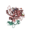
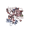
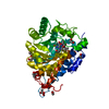
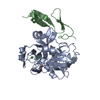
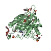
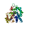
| Title | Sulphotransferase-18 from Arabidopsis thaliana in complex with 3'-phosphoadenosine 5'-phosphate (PAP)and sinigrin | ||||||||||||
 Components Components | Cytosolic sulfotransferase 18 | ||||||||||||
 Keywords Keywords | TRANSFERASE / Sulphotransferases / glucosinolate-biosynthesis / catalysis | ||||||||||||
| Function / homology |  Function and homology information Function and homology informationaliphatic desulfoglucosinolate sulfotransferase / aliphatic desulfoglucosinolate sulfotransferase activity / aromatic desulfoglucosinolate sulfotransferase activity / glucosinolate biosynthetic process / sulfation / cytoplasm Similarity search - Function | ||||||||||||
| Biological species |  | ||||||||||||
| Method |  X-RAY DIFFRACTION / SYNCHROTRON / MOLECULAR REPLACEMENT / Resolution: 1.92 Å X-RAY DIFFRACTION / SYNCHROTRON / MOLECULAR REPLACEMENT / Resolution: 1.92 Å | ||||||||||||
 Authors Authors | Hirschmann, F. / Krause, F. / Baruch, P. / Chizhov, I. / Mueller, J.W. / Manstein, D.J. / Papenbrock, J. / Fedorov, R. | ||||||||||||
| Funding support |  Germany, 2items Germany, 2items
| ||||||||||||
 Citation Citation | Journal: Sci Rep / Year: 2017 Title: Structural and biochemical studies of sulphotransferase 18 from Arabidopsis thaliana explain its substrate specificity and reaction mechanism. Authors: Hirschmann, F. / Krause, F. / Baruch, P. / Chizhov, I. / Mueller, J.W. / Manstein, D.J. / Papenbrock, J. / Fedorov, R. | ||||||||||||
| History |
|
- Structure visualization
| Structure viewer | Molecule: MolmilJmol/JSmol |
|---|
- Downloads & links
-Download
| PDBx/mmCIF format | 5mex.cif.gz | 91.7 KB | Display | PDBx/mmCIF format |
|---|---|---|---|---|
| PDB format | pdb5mex.ent.gz | 66.9 KB | Display | PDB format |
| PDBx/mmJSON format | 5mex.json.gz | Tree view | PDBx/mmJSON format | |
| Others |  Other downloads Other downloads |
-Validation report
| Arichive directory | https://data.pdbj.org/pub/pdb/validation_reports/me/5mexftp://data.pdbj.org/pub/pdb/validation_reports/me/5mex | HTTPS FTP |
|---|
-Related structure data
| Related structure data |  5mekC  1q44S S: Starting model for refinement C: citing same article ( |
|---|---|
| Similar structure data |


-Links
 PDBj
PDBj
- Assembly
| Deposited unit | 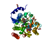
| ||||||||
|---|---|---|---|---|---|---|---|---|---|
| 1 |
| ||||||||
| Unit cell |
|
-Components
-Protein / Sugars , 2 types, 2 molecules A
| #1: Protein | Mass: 37499.508 Da / Num. of mol.: 1 Source method: isolated from a genetically manipulated source Source: (gene. exp.)  References: UniProt: Q9C9C9, Transferases; Transferring sulfur-containing groups; Sulfotransferases |
|---|---|
| #3: Sugar | ChemComp-SZZ /  Type: D-saccharide / Mass: 359.373 Da / Num. of mol.: 1 / Source method: obtained synthetically / Formula: C10H17NO9S2 Type: D-saccharide / Mass: 359.373 Da / Num. of mol.: 1 / Source method: obtained synthetically / Formula: C10H17NO9S2 |
-Non-polymers , 4 types, 298 molecules 


| #2: Chemical | ChemComp-PAP /  Mass: 507.181 Da / Num. of mol.: 1 / Source method: obtained synthetically / Formula: C10H16N5O13P3 Mass: 507.181 Da / Num. of mol.: 1 / Source method: obtained synthetically / Formula: C10H16N5O13P3 | ||||
|---|---|---|---|---|---|
| #4: Chemical | ChemComp-EDO /  Mass: 62.068 Da / Num. of mol.: 10 / Source method: obtained synthetically / Formula: C2H6O2 Mass: 62.068 Da / Num. of mol.: 10 / Source method: obtained synthetically / Formula: C2H6O2#5: Chemical | ChemComp-BU2 / |  Mass: 90.121 Da / Num. of mol.: 1 / Source method: obtained synthetically / Formula: C4H10O2 Mass: 90.121 Da / Num. of mol.: 1 / Source method: obtained synthetically / Formula: C4H10O2#6: Water | ChemComp-HOH / | Mass: 18.015 Da / Num. of mol.: 286 / Source method: isolated from a natural source / Formula: H2O |
-Experimental details
-Experiment
| Experiment | Method: X-RAY DIFFRACTION / Number of used crystals: 1 |
|---|
- Sample preparation
| Crystal | Density Matthews: 2.85 Å3/Da / Density % sol: 56.85 % |
|---|---|
| Crystal grow | Temperature: 291.15 K / Method: vapor diffusion / pH: 5.9 Details: 0.1 M 2-(N-morpholino) ethanesulfonic acid (MES) pH 5.9, 16% PEG4000, 160 mM NaCl, and 4% 1,3-butanediol |
-Data collection
| Diffraction | Mean temperature: 100 K |
|---|---|
| Diffraction source | Source: SYNCHROTRON / Site: ESRF  / Beamline: ID23-1 / Wavelength: 0.91 Å / Beamline: ID23-1 / Wavelength: 0.91 Å |
| Detector | Type: DECTRIS PILATUS 6M-F / Detector: PIXEL / Date: May 12, 2014 / Details: Pt coated mirrors in a Kirkpatrick-Baez geometry |
| Radiation | Monochromator: horizontally diffracting Si (111) monochromator Protocol: SINGLE WAVELENGTH / Monochromatic (M) / Laue (L): M / Scattering type: x-ray |
| Radiation wavelength | Wavelength: 0.91 Å / Relative weight: 1 |
| Reflection | Resolution: 1.92→47.16 Å / Num. obs: 34239 / % possible obs: 99.7 % / Redundancy: 10.55 % / Biso Wilson estimate: 37.25 Å2 / Rmerge(I) obs: 0.0647 / Rsym value: 0.027 / Net I/σ(I): 22.68 |
| Reflection shell | Resolution: 1.92→2.02 Å / Redundancy: 9.15 % / Rmerge(I) obs: 0.5435 / Mean I/σ(I) obs: 3.21 / % possible all: 98.3 |
- Processing
| Software |
| ||||||||||||||||||||
|---|---|---|---|---|---|---|---|---|---|---|---|---|---|---|---|---|---|---|---|---|---|
| Refinement | Method to determine structure: MOLECULAR REPLACEMENT Starting model: 1Q44 Resolution: 1.92→19.95 Å / Cor.coef. Fo:Fc: 0.969 / Cor.coef. Fo:Fc free: 0.943 / SU B: 3.105 / SU ML: 0.089 / Cross valid method: THROUGHOUT / ESU R: 0.128 / ESU R Free: 0.13 / Details: HYDROGENS HAVE BEEN ADDED IN THE RIDING POSITIONS
| ||||||||||||||||||||
| Solvent computation | Ion probe radii: 0.8 Å / Shrinkage radii: 0.8 Å / VDW probe radii: 1.2 Å | ||||||||||||||||||||
| Displacement parameters | Biso mean: 33.268 Å2
| ||||||||||||||||||||
| Refinement step | Cycle: 1 / Resolution: 1.92→19.95 Å
| ||||||||||||||||||||
| Refine LS restraints | Type: r_sphericity_bonded | ||||||||||||||||||||
| LS refinement shell | Resolution: 1.92→1.969 Å / Total num. of bins used: 20
|
