 Movie
Movie Controller


+ Open data

- Basic information
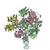
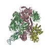
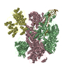
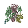
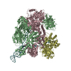
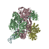
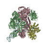
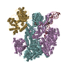
| Entry | Database: PDB / ID: 5mbv | ||||||
|---|---|---|---|---|---|---|---|
| Title | Cryo-EM structure of Lambda Phage protein GamS bound to RecBCD. | ||||||
 Components Components |
| ||||||
 Keywords Keywords | DNA BINDING PROTEIN / Inhibitor / Complex / DNA repair / Helicase/Nuclease | ||||||
| Function / homology |  Function and homology information Function and homology informationsymbiont-mediated evasion of DNA end degradation by host / deoxyribonuclease inhibitor activity / exodeoxyribonuclease V / exodeoxyribonuclease V activity / exodeoxyribonuclease V complex / clearance of foreign intracellular DNA / DNA translocase activity / DNA 5'-3' helicase / single-stranded DNA helicase activity / recombinational repair ...symbiont-mediated evasion of DNA end degradation by host / deoxyribonuclease inhibitor activity / exodeoxyribonuclease V / exodeoxyribonuclease V activity / exodeoxyribonuclease V complex / clearance of foreign intracellular DNA / DNA translocase activity / DNA 5'-3' helicase / single-stranded DNA helicase activity / recombinational repair / 3'-5' DNA helicase activity / DNA 3'-5' helicase / ATP-dependent activity, acting on DNA / DNA helicase activity / helicase activity / DNA endonuclease activity / response to radiation / double-strand break repair via homologous recombination / DNA recombination / 5'-3' DNA helicase activity / symbiont-mediated suppression of host innate immune response / DNA damage response / magnesium ion binding / ATP hydrolysis activity / DNA binding / ATP binding / cytosol Similarity search - Function | ||||||
| Biological species |   Enterobacteria phage lambda (virus) Enterobacteria phage lambda (virus) | ||||||
| Method | ELECTRON MICROSCOPY / single particle reconstruction / cryo EM / Resolution: 3.8 Å | ||||||
 Authors Authors | Wilkinson, M. / Chaban, Y. / Wigley, D.B. | ||||||
 Citation Citation | Journal: Elife / Year: 2016 Title: Structural basis for the inhibition of RecBCD by Gam and its synergistic antibacterial effect with quinolones. Authors: Martin Wilkinson / Luca Troman / Wan Ak Wan Nur Ismah / Yuriy Chaban / Matthew B Avison / Mark S Dillingham / Dale B Wigley /  Abstract: Our previous paper (Wilkinson , 2016) used high-resolution cryo-electron microscopy to solve the structure of the RecBCD complex, which acts in both the repair of double-stranded DNA breaks and the ...Our previous paper (Wilkinson , 2016) used high-resolution cryo-electron microscopy to solve the structure of the RecBCD complex, which acts in both the repair of double-stranded DNA breaks and the degradation of bacteriophage DNA. To counteract the latter activity, bacteriophage λ encodes a small protein inhibitor called Gam that binds to RecBCD and inactivates the complex. Here, we show that Gam inhibits RecBCD by competing at the DNA-binding site. The interaction surface is extensive and involves molecular mimicry of the DNA substrate. We also show that expression of Gam in or increases sensitivity to fluoroquinolones; antibacterials that kill cells by inhibiting topoisomerases and inducing double-stranded DNA breaks. Furthermore, fluoroquinolone-resistance in clinical isolates is reversed by expression of Gam. Together, our data explain the synthetic lethality observed between topoisomerase-induced DNA breaks and the RecBCD gene products, suggesting a new co-antibacterial strategy. | ||||||
| History |
|
- Structure visualization
| Movie |
Movie viewer |
|---|---|
| Structure viewer | Molecule: MolmilJmol/JSmol |
- Downloads & links
-Download
| PDBx/mmCIF format | 5mbv.cif.gz | 608.1 KB | Display | PDBx/mmCIF format |
|---|---|---|---|---|
| PDB format | pdb5mbv.ent.gz | 487.4 KB | Display | PDB format |
| PDBx/mmJSON format | 5mbv.json.gz | Tree view | PDBx/mmJSON format | |
| Others |  Other downloads Other downloads |
-Validation report
| Arichive directory | https://data.pdbj.org/pub/pdb/validation_reports/mb/5mbvftp://data.pdbj.org/pub/pdb/validation_reports/mb/5mbv | HTTPS FTP |
|---|
-Related structure data
| Related structure data |  3460MC M: map data used to model this data C: citing same article ( |
|---|---|
| Similar structure data |


-Links
 PDBj
PDBj


- Assembly
| Deposited unit | 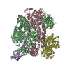
|
|---|---|
| 1 |
|
-Components
| #1: Protein | Mass: 134167.703 Da / Num. of mol.: 1 Source method: isolated from a genetically manipulated source Source: (gene. exp.) |
|---|---|
| #2: Protein | Mass: 128974.102 Da / Num. of mol.: 1 Source method: isolated from a genetically manipulated source Source: (gene. exp.) |
| #3: Protein | Mass: 67047.422 Da / Num. of mol.: 1 Source method: isolated from a genetically manipulated source Source: (gene. exp.) |
| #4: Protein | Mass: 11661.924 Da / Num. of mol.: 2 / Mutation: L79I same as crystal structure Source method: isolated from a genetically manipulated source Details: GamL is cleaved at position Y44 when expressed in E.coli into GamS (see paper - pubmed id 17544443). We had the GamS gene synthesised to start at M41 for this project. It purifies as a dimer. Source: (gene. exp.) Enterobacteria phage lambda (virus) / Gene: gam, gamma / Plasmid: pET22b / Production host: |
-Experimental details
-Experiment
| Experiment | Method: ELECTRON MICROSCOPY |
|---|---|
| EM experiment | Aggregation state: PARTICLE / 3D reconstruction method: single particle reconstruction |
- Sample preparation
| Component |
| ||||||||||||||||||||||||||||
|---|---|---|---|---|---|---|---|---|---|---|---|---|---|---|---|---|---|---|---|---|---|---|---|---|---|---|---|---|---|
| Molecular weight |
| ||||||||||||||||||||||||||||
| Source (natural) |
| ||||||||||||||||||||||||||||
| Source (recombinant) |
| ||||||||||||||||||||||||||||
| Buffer solution | pH: 7.5 | ||||||||||||||||||||||||||||
| Buffer component |
| ||||||||||||||||||||||||||||
| Specimen | Conc.: 1.4 mg/ml / Embedding applied: NO / Shadowing applied: NO / Staining applied: NO / Vitrification applied: YES | ||||||||||||||||||||||||||||
| Specimen support | Details: Grids were thinned by glow discharge in 30s steps with 1 minute wait in between treatments. Then left for 1-2 weeks prior to overnight treatment with 1 mM Ampiphol A8-35 to render surface ...Details: Grids were thinned by glow discharge in 30s steps with 1 minute wait in between treatments. Then left for 1-2 weeks prior to overnight treatment with 1 mM Ampiphol A8-35 to render surface hydrophilic. Grids were washed with 5 drops of water prior to use. EMS Grid mesh size: 400 divisions/in. / Grid type: C-flat-1/1 | ||||||||||||||||||||||||||||
| Vitrification | Instrument: FEI VITROBOT MARK IV / Cryogen name: ETHANE / Humidity: 90 % / Chamber temperature: 277 K / Details: 3 ul sample applied Blot force of -4 for 1 s |
- Electron microscopy imaging
| Experimental equipment |  Model: Titan Krios / Image courtesy: FEI Company |
|---|---|
| Microscopy | Model: FEI TITAN KRIOS |
| Electron gun | Electron source:  FIELD EMISSION GUN / Accelerating voltage: 300 kV / Illumination mode: FLOOD BEAM FIELD EMISSION GUN / Accelerating voltage: 300 kV / Illumination mode: FLOOD BEAM |
| Electron lens | Mode: BRIGHT FIELD / Nominal magnification: 105000 X / Calibrated magnification: 37313 X / Nominal defocus max: 2400 nm / Nominal defocus min: 1200 nm / Calibrated defocus min: 600 nm / Calibrated defocus max: 2900 nm / Cs: 2.7 mm / C2 aperture diameter: 70 µm / Alignment procedure: COMA FREE |
| Specimen holder | Cryogen: NITROGEN / Specimen holder model: FEI TITAN KRIOS AUTOGRID HOLDER |
| Image recording | Average exposure time: 0.4 sec. / Electron dose: 1.4 e/Å2 / Detector mode: COUNTING / Film or detector model: GATAN K2 SUMMIT (4k x 4k) / Num. of grids imaged: 1 / Num. of real images: 334 |
| Image scans | Sampling size: 5 µm / Movie frames/image: 25 / Used frames/image: 1-25 |
- Processing
| Software | Name: PHENIX / Version: 1.10.1_2155: / Classification: refinement | |||||||||||||||||||||||||||||||||||||||||||||||||||||||
|---|---|---|---|---|---|---|---|---|---|---|---|---|---|---|---|---|---|---|---|---|---|---|---|---|---|---|---|---|---|---|---|---|---|---|---|---|---|---|---|---|---|---|---|---|---|---|---|---|---|---|---|---|---|---|---|---|
| EM software |
| |||||||||||||||||||||||||||||||||||||||||||||||||||||||
| Image processing | Details: Frames were aligned using motioncorr prior to processing. | |||||||||||||||||||||||||||||||||||||||||||||||||||||||
| CTF correction | Details: CTF correction done at start of processing / Type: PHASE FLIPPING ONLY | |||||||||||||||||||||||||||||||||||||||||||||||||||||||
| Particle selection | Num. of particles selected: 134124 | |||||||||||||||||||||||||||||||||||||||||||||||||||||||
| Symmetry | Point symmetry: C1 (asymmetric) | |||||||||||||||||||||||||||||||||||||||||||||||||||||||
| 3D reconstruction | Resolution: 3.8 Å / Resolution method: FSC 0.143 CUT-OFF / Num. of particles: 122796 / Symmetry type: POINT | |||||||||||||||||||||||||||||||||||||||||||||||||||||||
| Atomic model building |
| |||||||||||||||||||||||||||||||||||||||||||||||||||||||
| Atomic model building |
| |||||||||||||||||||||||||||||||||||||||||||||||||||||||
| Refine LS restraints |
|

