 Movie
Movie Controller

[English] 日本語
 Yorodumi
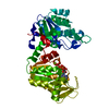
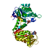
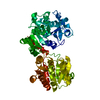
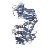
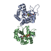
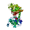
- PDB-5m6z: The X-ray structure of human M189I PGK-1 mutant in partially clos... -
+ Open data

- Basic information
| Entry | Database: PDB / ID: 5m6z | ||||||
|---|---|---|---|---|---|---|---|
| Title | The X-ray structure of human M189I PGK-1 mutant in partially closed conformation | ||||||
 Components Components | Phosphoglycerate kinase 1 | ||||||
 Keywords Keywords | TRANSFERASE / human phosphoglycerate kinase 1 / M189I SNP -derived mutant | ||||||
| Function / homology |  Function and homology information Function and homology informationnegative regulation of pyruvate decarboxylation to acetyl-CoA / Manipulation of host energy metabolism / phosphoglycerate kinase / phosphoglycerate kinase activity / protein-disulfide reductase [NAD(P)H] activity / Gluconeogenesis / Glycolysis / plasminogen activation / canonical glycolysis / epithelial cell differentiation ...negative regulation of pyruvate decarboxylation to acetyl-CoA / Manipulation of host energy metabolism / phosphoglycerate kinase / phosphoglycerate kinase activity / protein-disulfide reductase [NAD(P)H] activity / Gluconeogenesis / Glycolysis / plasminogen activation / canonical glycolysis / epithelial cell differentiation / negative regulation of angiogenesis / glycolytic process / gluconeogenesis / ADP binding / cellular response to hypoxia / transmembrane transporter binding / non-specific serine/threonine protein kinase / membrane raft / mitochondrial matrix / protein serine kinase activity / protein serine/threonine kinase activity / : / extracellular exosome / ATP binding / membrane / metal ion binding / cytosol Similarity search - Function | ||||||
| Biological species |  Homo sapiens (human) Homo sapiens (human) | ||||||
| Method |  X-RAY DIFFRACTION / SYNCHROTRON / MOLECULAR REPLACEMENT / Resolution: 1.67 Å X-RAY DIFFRACTION / SYNCHROTRON / MOLECULAR REPLACEMENT / Resolution: 1.67 Å | ||||||
 Authors Authors | Ilari, A. / Fiorillo, A. / Petrosino, M. / Cipollone, A. | ||||||
 Citation Citation | Journal: PLoS ONE / Year: 2018 Title: The phosphoglycerate kinase 1 variants found in carcinoma cells display different catalytic activity and conformational stability compared to the native enzyme. Authors: Fiorillo, A. / Petrosino, M. / Ilari, A. / Pasquo, A. / Cipollone, A. / Maggi, M. / Chiaraluce, R. / Consalvi, V. | ||||||
| History |
|
- Structure visualization
| Structure viewer | Molecule: MolmilJmol/JSmol |
|---|
- Downloads & links
-Download
| PDBx/mmCIF format | 5m6z.cif.gz | 99 KB | Display | PDBx/mmCIF format |
|---|---|---|---|---|
| PDB format | pdb5m6z.ent.gz | 72.2 KB | Display | PDB format |
| PDBx/mmJSON format | 5m6z.json.gz | Tree view | PDBx/mmJSON format | |
| Others |  Other downloads Other downloads |
-Validation report
| Arichive directory | https://data.pdbj.org/pub/pdb/validation_reports/m6/5m6zftp://data.pdbj.org/pub/pdb/validation_reports/m6/5m6z | HTTPS FTP |
|---|
-Related structure data
| Related structure data |  5m1rC 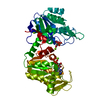 5m3uC 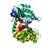 5mxmC 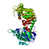 5o7dC 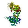 2zgvS S: Starting model for refinement C: citing same article ( |
|---|---|
| Similar structure data |


-Links
 PDBj
PDBj

- Assembly
| Deposited unit | 
| ||||||||
|---|---|---|---|---|---|---|---|---|---|
| 1 |
| ||||||||
| Unit cell |
|
-Components
-Protein , 1 types, 1 molecules A
| #1: Protein | Mass: 44523.387 Da / Num. of mol.: 1 / Mutation: M189I Source method: isolated from a genetically manipulated source Source: (gene. exp.) Homo sapiens (human) / Gene: PGK1, PGKA, MIG10, OK/SW-cl.110 / Production host:  |
|---|
-Non-polymers , 5 types, 205 molecules 


| #2: Chemical | ChemComp-3PG /  Mass: 186.057 Da / Num. of mol.: 1 / Source method: obtained synthetically / Formula: C3H7O7P Mass: 186.057 Da / Num. of mol.: 1 / Source method: obtained synthetically / Formula: C3H7O7P | ||
|---|---|---|---|
| #3: Chemical | ChemComp-PO4 /  Mass: 94.971 Da / Num. of mol.: 1 / Source method: obtained synthetically / Formula: PO4 Mass: 94.971 Da / Num. of mol.: 1 / Source method: obtained synthetically / Formula: PO4 | ||
| #4: Chemical | ChemComp-ADP /  Mass: 427.201 Da / Num. of mol.: 1 / Source method: obtained synthetically / Formula: C10H15N5O10P2 / Comment: ADP, energy-carrying molecule*YM Mass: 427.201 Da / Num. of mol.: 1 / Source method: obtained synthetically / Formula: C10H15N5O10P2 / Comment: ADP, energy-carrying molecule*YM | ||
| #5: Chemical |  Mass: 24.305 Da / Num. of mol.: 2 / Source method: obtained synthetically / Formula: Mg Mass: 24.305 Da / Num. of mol.: 2 / Source method: obtained synthetically / Formula: Mg#6: Water | ChemComp-HOH / | Mass: 18.015 Da / Num. of mol.: 200 / Source method: isolated from a natural source / Formula: H2O |
-Experimental details
-Experiment
| Experiment | Method: X-RAY DIFFRACTION / Number of used crystals: 1 |
|---|
- Sample preparation
| Crystal | Density Matthews: 2.02 Å3/Da / Density % sol: 38.51 % |
|---|---|
| Crystal grow | Temperature: 294 K / Method: vapor diffusion, hanging drop / pH: 8.6 / Details: 2.5 M Na//KPO4, pH=8.6 |
-Data collection
| Diffraction | Mean temperature: 100 K |
|---|---|
| Diffraction source | Source: SYNCHROTRON / Site: ESRF  / Beamline: ID23-2 / Wavelength: 0.872899 Å / Beamline: ID23-2 / Wavelength: 0.872899 Å |
| Detector | Type: DECTRIS PILATUS3 2M / Detector: PIXEL / Date: May 13, 2016 |
| Radiation | Protocol: SINGLE WAVELENGTH / Monochromatic (M) / Laue (L): M / Scattering type: x-ray |
| Radiation wavelength | Wavelength: 0.872899 Å / Relative weight: 1 |
| Reflection | Resolution: 1.67→60 Å / Num. obs: 42601 / % possible obs: 97.5 % / Redundancy: 4.26 % / CC1/2: 0.99 / Rmerge(I) obs: 0.054 / Net I/σ(I): 15.08 |
| Reflection shell | Resolution: 1.67→1.77 Å / Redundancy: 4.18 % / Rmerge(I) obs: 0.675 / Mean I/σ(I) obs: 1.89 / CC1/2: 0.59 / % possible all: 94.1 |
- Processing
| Software |
| ||||||||||||||||||||||||||||||||||||||||||||||||||||||||||||||||||||||||||||||||||||||||||||||||||||||||||||||||||||||||||||||||||||||||||||||||||||||||||||||||||||||||||||||||||||||
|---|---|---|---|---|---|---|---|---|---|---|---|---|---|---|---|---|---|---|---|---|---|---|---|---|---|---|---|---|---|---|---|---|---|---|---|---|---|---|---|---|---|---|---|---|---|---|---|---|---|---|---|---|---|---|---|---|---|---|---|---|---|---|---|---|---|---|---|---|---|---|---|---|---|---|---|---|---|---|---|---|---|---|---|---|---|---|---|---|---|---|---|---|---|---|---|---|---|---|---|---|---|---|---|---|---|---|---|---|---|---|---|---|---|---|---|---|---|---|---|---|---|---|---|---|---|---|---|---|---|---|---|---|---|---|---|---|---|---|---|---|---|---|---|---|---|---|---|---|---|---|---|---|---|---|---|---|---|---|---|---|---|---|---|---|---|---|---|---|---|---|---|---|---|---|---|---|---|---|---|---|---|---|---|
| Refinement | Method to determine structure: MOLECULAR REPLACEMENT Starting model: 2ZGV Resolution: 1.67→53.06 Å / Cor.coef. Fo:Fc: 0.968 / Cor.coef. Fo:Fc free: 0.949 / SU B: 2.867 / SU ML: 0.092 / Cross valid method: THROUGHOUT / ESU R: 0.111 / ESU R Free: 0.11 / Details: HYDROGENS HAVE BEEN ADDED IN THE RIDING POSITIONS
| ||||||||||||||||||||||||||||||||||||||||||||||||||||||||||||||||||||||||||||||||||||||||||||||||||||||||||||||||||||||||||||||||||||||||||||||||||||||||||||||||||||||||||||||||||||||
| Solvent computation | Ion probe radii: 0.8 Å / Shrinkage radii: 0.8 Å / VDW probe radii: 1.2 Å | ||||||||||||||||||||||||||||||||||||||||||||||||||||||||||||||||||||||||||||||||||||||||||||||||||||||||||||||||||||||||||||||||||||||||||||||||||||||||||||||||||||||||||||||||||||||
| Displacement parameters | Biso mean: 27.804 Å2
| ||||||||||||||||||||||||||||||||||||||||||||||||||||||||||||||||||||||||||||||||||||||||||||||||||||||||||||||||||||||||||||||||||||||||||||||||||||||||||||||||||||||||||||||||||||||
| Refinement step | Cycle: 1 / Resolution: 1.67→53.06 Å
| ||||||||||||||||||||||||||||||||||||||||||||||||||||||||||||||||||||||||||||||||||||||||||||||||||||||||||||||||||||||||||||||||||||||||||||||||||||||||||||||||||||||||||||||||||||||
| Refine LS restraints |
|