 Movie
Movie Controller


+ Open data

- Basic information
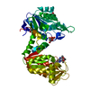
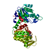
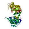
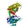
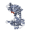
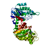
| Entry | Database: PDB / ID: 1fw8 | ||||||
|---|---|---|---|---|---|---|---|
| Title | CIRCULARLY PERMUTED PHOSPHOGLYCERATE KINASE FROM YEAST: PGK P72 | ||||||
 Components Components | Phosphoglycerate kinase | ||||||
 Keywords Keywords | TRANSFERASE / phosphotransferase / kinase / phosphoglycerate kinase / glycolysis / mutant / permutation / permuted sequence / PGK / protein folding / two-domain protein | ||||||
| Function / homology |  Function and homology information Function and homology informationGluconeogenesis / Glycolysis / phosphoglycerate kinase / phosphoglycerate kinase activity / glycolytic process / gluconeogenesis / ADP binding / mitochondrion / ATP binding / metal ion binding ...Gluconeogenesis / Glycolysis / phosphoglycerate kinase / phosphoglycerate kinase activity / glycolytic process / gluconeogenesis / ADP binding / mitochondrion / ATP binding / metal ion binding / plasma membrane / cytoplasm / cytosol Similarity search - Function | ||||||
| Biological species |  | ||||||
| Method |  X-RAY DIFFRACTION / SYNCHROTRON / Resolution: 2.3 Å X-RAY DIFFRACTION / SYNCHROTRON / Resolution: 2.3 Å | ||||||
 Authors Authors | Tougard, P. / Bizebard, T. / Ritco-Vonsovici, M. / Minard, P. / Desmadril, M. | ||||||
 Citation Citation | Journal: Acta Crystallogr.,Sect.D / Year: 2002 Title: Structure of a circularly permuted phosphoglycerate kinase. Authors: Tougard, P. / Bizebard, T. / Ritco-Vonsovici, M. / Minard, P. / Desmadril, M. #1: Journal: Biochemistry / Year: 1995Title: Is the continuity of the domains required for the correct folding of a two-domain protein? Authors: Ritco-Vonsovici, M. / Minard, P. / Desmadril, M. / Yon, J.M. | ||||||
| History |
|
- Structure visualization
| Structure viewer | Molecule: MolmilJmol/JSmol |
|---|
- Downloads & links
-Download
| PDBx/mmCIF format | 1fw8.cif.gz | 95.3 KB | Display | PDBx/mmCIF format |
|---|---|---|---|---|
| PDB format | pdb1fw8.ent.gz | 71.5 KB | Display | PDB format |
| PDBx/mmJSON format | 1fw8.json.gz | Tree view | PDBx/mmJSON format | |
| Others |  Other downloads Other downloads |
-Validation report
| Arichive directory | https://data.pdbj.org/pub/pdb/validation_reports/fw/1fw8ftp://data.pdbj.org/pub/pdb/validation_reports/fw/1fw8 | HTTPS FTP |
|---|
-Related structure data
| Similar structure data |
|---|


-Links
 PDBj
PDBj
- Assembly
| Deposited unit | 
| ||||||||
|---|---|---|---|---|---|---|---|---|---|
| 1 |
| ||||||||
| Unit cell |
|
-Components
| #1: Protein | Mass: 44654.121 Da / Num. of mol.: 1 Source method: isolated from a genetically manipulated source Source: (gene. exp.) Strain: ATCC 204508 / S288c / Gene: PGK1, YCR012W, YCR12W / Plasmid: PYE / Production host: | ||||
|---|---|---|---|---|---|
| #2: Chemical | ChemComp-GOL /   Mass: 92.094 Da / Num. of mol.: 4 / Source method: obtained synthetically / Formula: C3H8O3 Mass: 92.094 Da / Num. of mol.: 4 / Source method: obtained synthetically / Formula: C3H8O3#3: Water | ChemComp-HOH / |  Mass: 18.015 Da / Num. of mol.: 181 / Source method: isolated from a natural source / Formula: H2O Mass: 18.015 Da / Num. of mol.: 181 / Source method: isolated from a natural source / Formula: H2OHas protein modification | Y | |
-Experimental details
-Experiment
| Experiment | Method: X-RAY DIFFRACTION / Number of used crystals: 1 |
|---|
- Sample preparation
| Crystal | Density Matthews: 2.23 Å3/Da / Density % sol: 44.82 % | |||||||||||||||
|---|---|---|---|---|---|---|---|---|---|---|---|---|---|---|---|---|
| Crystal grow | Temperature: 293 K / Method: vapor diffusion, hanging drop / pH: 6.1 Details: ammonium sulfate, sodium pyrophosphate, dioxan, glycerol for cryogenic conditions, pH 6.1, VAPOR DIFFUSION, HANGING DROP, temperature 293K | |||||||||||||||
| Crystal grow | *PLUS | |||||||||||||||
| Components of the solutions | *PLUS
|
-Data collection
| Diffraction | Mean temperature: 120 K |
|---|---|
| Diffraction source | Source: SYNCHROTRON / Site: LURE  / Beamline: D41A / Wavelength: 1.375 / Beamline: D41A / Wavelength: 1.375 |
| Detector | Type: MARRESEARCH / Detector: IMAGE PLATE / Date: Sep 10, 1997 |
| Radiation | Protocol: SINGLE WAVELENGTH / Monochromatic (M) / Laue (L): M / Scattering type: x-ray |
| Radiation wavelength | Wavelength: 1.375 Å / Relative weight: 1 |
| Reflection | Resolution: 2.3→14.99 Å / Num. all: 18305 / % possible obs: 99.8 % / Redundancy: 3.5 % / Rmerge(I) obs: 0.079 |
| Reflection shell | Resolution: 2.3→2.8 Å / Redundancy: 3.3 % / Rmerge(I) obs: 0.273 / Num. unique all: 1787 / % possible all: 99.4 |
| Reflection | *PLUS Num. obs: 18306 / Num. measured all: 64243 |
| Reflection shell | *PLUS % possible obs: 99.4 % / Rmerge(I) obs: 0.322 / Mean I/σ(I) obs: 3.5 |
- Processing
| Software |
| ||||||||||||||||
|---|---|---|---|---|---|---|---|---|---|---|---|---|---|---|---|---|---|
| Refinement | Resolution: 2.3→7 Å
| ||||||||||||||||
| Refinement step | Cycle: LAST / Resolution: 2.3→7 Å
| ||||||||||||||||
| Refine LS restraints |
| ||||||||||||||||
| Software | *PLUS Name: X-PLOR / Version: 3.1 / Classification: refinement | ||||||||||||||||
| Refinement | *PLUS σ(I): 16817 | ||||||||||||||||
| Solvent computation | *PLUS | ||||||||||||||||
| Displacement parameters | *PLUS | ||||||||||||||||
| Refine LS restraints | *PLUS
|
