 Movie
Movie Controller

[English] 日本語
 Yorodumi
- PDB-5m58: Crystal structure of CouO, a C-methyltransferase from Streptomyce... -
+ Open data

- Basic information
| Entry | Database: PDB / ID: 5m58 | ||||||
|---|---|---|---|---|---|---|---|
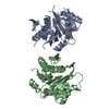
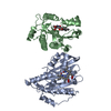
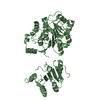
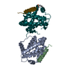
| Title | Crystal structure of CouO, a C-methyltransferase from Streptomyces rishiriensis | ||||||
 Components Components | C-methyltransferase CouO | ||||||
 Keywords Keywords | TRANSFERASE / C-methyltransferase / CouO / Friedel-Craft alkylation / SAM / transfrease | ||||||
| Function / homology | Methyltransferase domain 25 / Methyltransferase domain / antibiotic biosynthetic process / Transferases; Transferring one-carbon groups; Methyltransferases / methyltransferase activity / methylation / S-adenosyl-L-methionine-dependent methyltransferase superfamily / S-ADENOSYL-L-HOMOCYSTEINE / C-methyltransferase CouO Function and homology information Function and homology information | ||||||
| Biological species |  Streptomyces rishiriensis (bacteria) Streptomyces rishiriensis (bacteria) | ||||||
| Method |  X-RAY DIFFRACTION / SYNCHROTRON / MOLECULAR REPLACEMENT / Resolution: 2.05 Å X-RAY DIFFRACTION / SYNCHROTRON / MOLECULAR REPLACEMENT / Resolution: 2.05 Å | ||||||
 Authors Authors | Pavkov-Keller, T. / Gruber, K. | ||||||
 Citation Citation | Journal: PLoS ONE / Year: 2017 Title: Crystal Structure and Catalytic Mechanism of CouO, a Versatile C-Methyltransferase from Streptomyces rishiriensis. Authors: Pavkov-Keller, T. / Steiner, K. / Faber, M. / Tengg, M. / Schwab, H. / Gruber-Khadjawi, M. / Gruber, K. #1: Journal: Acta Crystallogr. Sect. F Struct. Biol. Cryst. Commun. Year: 2012 Title: Crystallization of the novel S-adenosyl-L-methionine-dependent C-methyltransferase CouO from Streptomyces rishiriensis and preliminary diffraction data analysis. Authors: Lyskowski, A. / Tengg, M. / Steinkellner, G. / Schwab, H. / Gruber-Khadjawi, M. / Gruber, K. | ||||||
| History |
|
- Structure visualization
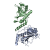
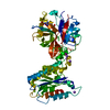
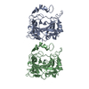
| Structure viewer | Molecule: MolmilJmol/JSmol |
|---|
- Downloads & links
-Download
| PDBx/mmCIF format | 5m58.cif.gz | 110.6 KB | Display | PDBx/mmCIF format |
|---|---|---|---|---|
| PDB format | pdb5m58.ent.gz | 84.6 KB | Display | PDB format |
| PDBx/mmJSON format | 5m58.json.gz | Tree view | PDBx/mmJSON format | |
| Others |  Other downloads Other downloads |
-Validation report
| Arichive directory | https://data.pdbj.org/pub/pdb/validation_reports/m5/5m58ftp://data.pdbj.org/pub/pdb/validation_reports/m5/5m58 | HTTPS FTP |
|---|
-Related structure data
| Similar structure data |
|---|

-Links
 PDBj
PDBj- Assembly
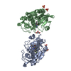
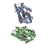
| Deposited unit | 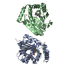
| ||||||||
|---|---|---|---|---|---|---|---|---|---|
| 1 |
| ||||||||
| Unit cell |
|
-Components
| #1: Protein | Mass: 25680.061 Da / Num. of mol.: 2 Source method: isolated from a genetically manipulated source Source: (gene. exp.) Streptomyces rishiriensis (bacteria) / Gene: couO / Production host: References: UniProt: Q9F8T9, Transferases; Transferring one-carbon groups; Methyltransferases #2: Chemical |   Mass: 384.411 Da / Num. of mol.: 2 / Source method: obtained synthetically / Formula: C14H20N6O5S Mass: 384.411 Da / Num. of mol.: 2 / Source method: obtained synthetically / Formula: C14H20N6O5S#3: Water | ChemComp-HOH / |  Mass: 18.015 Da / Num. of mol.: 314 / Source method: isolated from a natural source / Formula: H2O Mass: 18.015 Da / Num. of mol.: 314 / Source method: isolated from a natural source / Formula: H2OHas protein modification | N | |
|---|
-Experimental details
-Experiment
| Experiment | Method: X-RAY DIFFRACTION / Number of used crystals: 1 |
|---|
- Sample preparation
| Crystal | Density Matthews: 2.04 Å3/Da / Density % sol: 39.65 % |
|---|---|
| Crystal grow | Temperature: 290 K / Method: vapor diffusion, sitting drop Details: 2.3 mg/ml CouO 12.5%(w/v) PEG 1000, 12.5%(w/v) PEG 3350, 12.5%(v/v) MPD, 0.03 M of each halide (sodium fluoride, sodium bromide and sodium iodide), 0.1 M MES/imidazole pH 6.5 |
-Data collection
| Diffraction | Mean temperature: 100 K |
|---|---|
| Diffraction source | Source: SYNCHROTRON / Site: EMBL/DESY, HAMBURG  / Beamline: X12 / Wavelength: 1.0507 Å / Beamline: X12 / Wavelength: 1.0507 Å |
| Detector | Type: MARRESEARCH / Detector: CCD / Date: Oct 13, 2011 |
| Radiation | Protocol: SINGLE WAVELENGTH / Monochromatic (M) / Laue (L): M / Scattering type: x-ray |
| Radiation wavelength | Wavelength: 1.0507 Å / Relative weight: 1 |
| Reflection | Resolution: 2.05→40.98 Å / Num. all: 87179 / Num. obs: 25206 / % possible obs: 97.9 % / Redundancy: 3.5 % / Rmerge(I) obs: 0.13 / Net I/σ(I): 6 |
| Reflection shell | Resolution: 2.05→2.16 Å / Redundancy: 3 % / Rmerge(I) obs: 0.25 / Mean I/σ(I) obs: 2.5 / Num. measured obs: 10510 / Num. unique all: 3498 / % possible all: 94.2 |
- Processing
| Software |
| ||||||||||||||||||||||||||||||||||||||||||||||||||||||||||||||||||||||||||||||||||||||||||||||||||||||||||||||||||||||||||||||||||||||||||||||||||||||||||||||||||||||||||||||||||||||
|---|---|---|---|---|---|---|---|---|---|---|---|---|---|---|---|---|---|---|---|---|---|---|---|---|---|---|---|---|---|---|---|---|---|---|---|---|---|---|---|---|---|---|---|---|---|---|---|---|---|---|---|---|---|---|---|---|---|---|---|---|---|---|---|---|---|---|---|---|---|---|---|---|---|---|---|---|---|---|---|---|---|---|---|---|---|---|---|---|---|---|---|---|---|---|---|---|---|---|---|---|---|---|---|---|---|---|---|---|---|---|---|---|---|---|---|---|---|---|---|---|---|---|---|---|---|---|---|---|---|---|---|---|---|---|---|---|---|---|---|---|---|---|---|---|---|---|---|---|---|---|---|---|---|---|---|---|---|---|---|---|---|---|---|---|---|---|---|---|---|---|---|---|---|---|---|---|---|---|---|---|---|---|---|
| Refinement | Method to determine structure: MOLECULAR REPLACEMENT / Resolution: 2.05→40 Å / Cor.coef. Fo:Fc: 0.931 / Cor.coef. Fo:Fc free: 0.869 / SU B: 6.833 / SU ML: 0.181 / Cross valid method: THROUGHOUT / ESU R: 0.295 / ESU R Free: 0.225 / Details: HYDROGENS HAVE BEEN ADDED IN THE RIDING POSITIONS
| ||||||||||||||||||||||||||||||||||||||||||||||||||||||||||||||||||||||||||||||||||||||||||||||||||||||||||||||||||||||||||||||||||||||||||||||||||||||||||||||||||||||||||||||||||||||
| Solvent computation | Ion probe radii: 0.8 Å / Shrinkage radii: 0.8 Å / VDW probe radii: 1.2 Å | ||||||||||||||||||||||||||||||||||||||||||||||||||||||||||||||||||||||||||||||||||||||||||||||||||||||||||||||||||||||||||||||||||||||||||||||||||||||||||||||||||||||||||||||||||||||
| Displacement parameters | Biso mean: 22.234 Å2
| ||||||||||||||||||||||||||||||||||||||||||||||||||||||||||||||||||||||||||||||||||||||||||||||||||||||||||||||||||||||||||||||||||||||||||||||||||||||||||||||||||||||||||||||||||||||
| Refinement step | Cycle: 1 / Resolution: 2.05→40 Å
| ||||||||||||||||||||||||||||||||||||||||||||||||||||||||||||||||||||||||||||||||||||||||||||||||||||||||||||||||||||||||||||||||||||||||||||||||||||||||||||||||||||||||||||||||||||||
| Refine LS restraints |
|