 Movie
Movie Controller

[English] 日本語
 Yorodumi
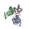
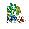
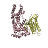
- PDB-5m4y: Crystal structure of the Sec3/Sso2 complex at 2.20 angstrom resolution -
+ Open data

- Basic information
| Entry | Database: PDB / ID: 5m4y | |||||||||
|---|---|---|---|---|---|---|---|---|---|---|
| Title | Crystal structure of the Sec3/Sso2 complex at 2.20 angstrom resolution | |||||||||
 Components Components |
| |||||||||
 Keywords Keywords | STRUCTURAL PROTEIN / exocyst / Sec3 / Sso2 | |||||||||
| Function / homology |  Function and homology information Function and homology informationInsulin processing / exocyst assembly / exocyst localization / endoplasmic reticulum inheritance / Disinhibition of SNARE formation / vesicle fusion to plasma membrane / ascospore-type prospore assembly / exocyst / Golgi vesicle fusion to target membrane / Insertion of tail-anchored proteins into the endoplasmic reticulum membrane ...Insulin processing / exocyst assembly / exocyst localization / endoplasmic reticulum inheritance / Disinhibition of SNARE formation / vesicle fusion to plasma membrane / ascospore-type prospore assembly / exocyst / Golgi vesicle fusion to target membrane / Insertion of tail-anchored proteins into the endoplasmic reticulum membrane / trans-Golgi Network Vesicle Budding / prospore membrane / COPII-mediated vesicle transport / incipient cellular bud site / : / cellular bud tip / : / SNARE complex / SNAP receptor activity / Golgi to plasma membrane transport / vesicle fusion / cellular bud neck / phosphatidic acid binding / mating projection tip / exocytosis / phosphatidylinositol-4,5-bisphosphate binding / endomembrane system / cell periphery / SNARE binding / intracellular protein transport / small GTPase binding / protein transport / Golgi membrane / endoplasmic reticulum / plasma membrane / cytoplasm Similarity search - Function | |||||||||
| Biological species |  | |||||||||
| Method |  X-RAY DIFFRACTION / SYNCHROTRON / MOLECULAR REPLACEMENT / Resolution: 2.2 Å X-RAY DIFFRACTION / SYNCHROTRON / MOLECULAR REPLACEMENT / Resolution: 2.2 Å | |||||||||
 Authors Authors | Zhang, Y.B. / Dong, G. | |||||||||
| Funding support |  Austria, 1items Austria, 1items
| |||||||||
 Citation Citation | Journal: Nat Commun / Year: 2017 Title: Sec3 promotes the initial binary t-SNARE complex assembly and membrane fusion. Authors: Yue, P. / Zhang, Y. / Mei, K. / Wang, S. / Lesigang, J. / Zhu, Y. / Dong, G. / Guo, W. | |||||||||
| History |
|
- Structure visualization
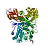
| Structure viewer | Molecule: MolmilJmol/JSmol |
|---|
- Downloads & links
-Download
| PDBx/mmCIF format | 5m4y.cif.gz | 233 KB | Display | PDBx/mmCIF format |
|---|---|---|---|---|
| PDB format | pdb5m4y.ent.gz | 185.1 KB | Display | PDB format |
| PDBx/mmJSON format | 5m4y.json.gz | Tree view | PDBx/mmJSON format | |
| Others |  Other downloads Other downloads |
-Validation report
| Arichive directory | https://data.pdbj.org/pub/pdb/validation_reports/m4/5m4yftp://data.pdbj.org/pub/pdb/validation_reports/m4/5m4y | HTTPS FTP |
|---|
-Related structure data
| Related structure data | 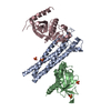 5lg4C  1ifoS 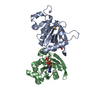 3a58S S: Starting model for refinement C: citing same article ( |
|---|---|
| Similar structure data |

-Links
 PDBj
PDBj

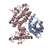
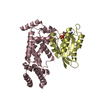
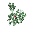
- Assembly
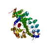
| Deposited unit | 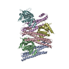
| ||||||||
|---|---|---|---|---|---|---|---|---|---|
| 1 | 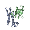
| ||||||||
| 2 | 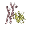
| ||||||||
| 3 | 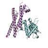
| ||||||||
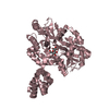
| Unit cell |
| ||||||||
| Components on special symmetry positions |
|
-Components
| #1: Protein | Mass: 24394.135 Da / Num. of mol.: 3 Source method: isolated from a genetically manipulated source Source: (gene. exp.) Gene: SSO2, YMR183C, YM8010.13C / Production host: Escherichia coli BL21(DE3) / References: UniProt: P39926 #2: Protein | Mass: 28513.105 Da / Num. of mol.: 3 Source method: isolated from a genetically manipulated source Source: (gene. exp.) Gene: SEC3, PSL1, YER008C / Production host:  #3: Chemical | ChemComp-GOL / |   Mass: 92.094 Da / Num. of mol.: 1 / Source method: obtained synthetically / Formula: C3H8O3 Mass: 92.094 Da / Num. of mol.: 1 / Source method: obtained synthetically / Formula: C3H8O3#4: Water | ChemComp-HOH / |  Mass: 18.015 Da / Num. of mol.: 325 / Source method: isolated from a natural source / Formula: H2O Mass: 18.015 Da / Num. of mol.: 325 / Source method: isolated from a natural source / Formula: H2O |
|---|
-Experimental details
-Experiment
| Experiment | Method: X-RAY DIFFRACTION / Number of used crystals: 1 |
|---|
- Sample preparation
| Crystal | Density Matthews: 2.01 Å3/Da / Density % sol: 38.78 % |
|---|---|
| Crystal grow | Temperature: 295 K / Method: vapor diffusion, sitting drop / pH: 7 Details: 0.1 M HEPES (pH 7.0), 0.2 M NaCl, and 25% (w/v) PEG 3350 |
-Data collection
| Diffraction | Mean temperature: 100 K |
|---|---|
| Diffraction source | Source: SYNCHROTRON / Site: ESRF  / Beamline: ID29 / Wavelength: 0.9793 Å / Beamline: ID29 / Wavelength: 0.9793 Å |
| Detector | Type: DECTRIS PILATUS 6M-F / Detector: PIXEL / Date: Dec 14, 2013 |
| Radiation | Protocol: SINGLE WAVELENGTH / Monochromatic (M) / Laue (L): M / Scattering type: x-ray |
| Radiation wavelength | Wavelength: 0.9793 Å / Relative weight: 1 |
| Reflection | Resolution: 2.2→50 Å / Num. obs: 62754 / % possible obs: 99.4 % / Redundancy: 6.5 % / Net I/σ(I): 13.2 |
- Processing
| Software |
| |||||||||||||||||||||||||||||||||||||||||||||||||||||||||||||||||||||||||||||||||||||||||||
|---|---|---|---|---|---|---|---|---|---|---|---|---|---|---|---|---|---|---|---|---|---|---|---|---|---|---|---|---|---|---|---|---|---|---|---|---|---|---|---|---|---|---|---|---|---|---|---|---|---|---|---|---|---|---|---|---|---|---|---|---|---|---|---|---|---|---|---|---|---|---|---|---|---|---|---|---|---|---|---|---|---|---|---|---|---|---|---|---|---|---|---|---|
| Refinement | Method to determine structure: MOLECULAR REPLACEMENT Starting model: 3A58, 1IFO Resolution: 2.2→19.978 Å / SU ML: 0.31 / Cross valid method: FREE R-VALUE / σ(F): 1.34 / Phase error: 28.83
| |||||||||||||||||||||||||||||||||||||||||||||||||||||||||||||||||||||||||||||||||||||||||||
| Solvent computation | Shrinkage radii: 0.9 Å / VDW probe radii: 1.11 Å | |||||||||||||||||||||||||||||||||||||||||||||||||||||||||||||||||||||||||||||||||||||||||||
| Refinement step | Cycle: LAST / Resolution: 2.2→19.978 Å
| |||||||||||||||||||||||||||||||||||||||||||||||||||||||||||||||||||||||||||||||||||||||||||
| Refine LS restraints |
| |||||||||||||||||||||||||||||||||||||||||||||||||||||||||||||||||||||||||||||||||||||||||||
| LS refinement shell |
|
