 Movie
Movie Controller

[English] 日本語
 Yorodumi
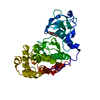
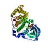
- PDB-5m4u: ORTHORHOMBIC COMPLEX STRUCTURE OF HUMAN PROTEIN KINASE CK2 CATALY... -
+ Open data

- Basic information
| Entry | Database: PDB / ID: 5m4u | |||||||||
|---|---|---|---|---|---|---|---|---|---|---|
| Title | ORTHORHOMBIC COMPLEX STRUCTURE OF HUMAN PROTEIN KINASE CK2 CATALYTIC SUBUNIT (ISOFORM CK2ALPHA') WITH THE INHIBITOR 4'-CARBOXY-6,8-CHLORO- FLAVONOL (FLC21) | |||||||||
 Components Components | Casein kinase II subunit alpha' | |||||||||
 Keywords Keywords | TRANSFERASE / protein kinase CK2 / casein kinase 2 | |||||||||
| Function / homology |  Function and homology information Function and homology informationSPOP-mediated proteasomal degradation of PD-L1(CD274) / regulation of mitophagy / regulation of chromosome separation / WNT mediated activation of DVL / Condensation of Prometaphase Chromosomes / protein kinase CK2 complex / : / Phosphorylation and nuclear translocation of the CRY:PER:kinase complex / Regulation of CDH1 posttranslational processing and trafficking to plasma membrane / Receptor Mediated Mitophagy ...SPOP-mediated proteasomal degradation of PD-L1(CD274) / regulation of mitophagy / regulation of chromosome separation / WNT mediated activation of DVL / Condensation of Prometaphase Chromosomes / protein kinase CK2 complex / : / Phosphorylation and nuclear translocation of the CRY:PER:kinase complex / Regulation of CDH1 posttranslational processing and trafficking to plasma membrane / Receptor Mediated Mitophagy / Synthesis of PC / RUNX1 interacts with co-factors whose precise effect on RUNX1 targets is not known / Maturation of hRSV A proteins / negative regulation of apoptotic signaling pathway / negative regulation of proteasomal ubiquitin-dependent protein catabolic process / liver regeneration / acrosomal vesicle / Signal transduction by L1 / cerebral cortex development / Wnt signaling pathway / Regulation of PTEN stability and activity / KEAP1-NFE2L2 pathway / double-strand break repair / Cooperation of PDCL (PhLP1) and TRiC/CCT in G-protein beta folding / heterochromatin formation / spermatogenesis / Regulation of TP53 Activity through Phosphorylation / non-specific serine/threonine protein kinase / protein serine kinase activity / protein serine/threonine kinase activity / apoptotic process / DNA damage response / positive regulation of DNA-templated transcription / chromatin / DNA-templated transcription / nucleoplasm / ATP binding / nucleus / cytosol Similarity search - Function | |||||||||
| Biological species |  Homo sapiens (human) Homo sapiens (human) | |||||||||
| Method |  X-RAY DIFFRACTION / SYNCHROTRON / MOLECULAR REPLACEMENT / Resolution: 2.195 Å X-RAY DIFFRACTION / SYNCHROTRON / MOLECULAR REPLACEMENT / Resolution: 2.195 Å | |||||||||
 Authors Authors | Niefind, K. / Bischoff, N. / Yarmoluk, S.M. / Bdzhola, V.G. / Golub, A.G. / Balanda, A.O. / Prykhod'ko, A.O. | |||||||||
| Funding support |  Germany, 1items Germany, 1items
| |||||||||
 Citation Citation | Journal: Pharmaceuticals / Year: 2017 Title: Structural Hypervariability of the Two Human Protein Kinase CK2 Catalytic Subunit Paralogs Revealed by Complex Structures with a Flavonol- and a Thieno[2,3-d]pyrimidine-Based Inhibitor. Authors: Niefind, K. / Bischoff, N. / Golub, A.G. / Bdzhola, V.G. / Balanda, A.O. / Prykhod'ko, A.O. / Yarmoluk, S.M. #1: Journal: ACS Chem. Biol. / Year: 2015Title: A Note of Caution on the Role of Halogen Bonds for Protein Kinase/Inhibitor Recognition Suggested by High- And Low-Salt CK2alpha Complex Structures. Authors: Guerra, B. / Bischoff, N. / Bdzhola, V.G. / Yarmoluk, S.M. / Issinger, O.G. / Golub, A.G. / Niefind, K. #2: Journal: Eur J Med Chem / Year: 2011 Title: Synthesis and biological evaluation of substituted (thieno[2,3-d]pyrimidin-4-ylthio)carboxylic acids as inhibitors of human protein kinase CK2. Authors: Golub, A.G. / Bdzhola, V.G. / Briukhovetska, N.V. / Balanda, A.O. / Kukharenko, O.P. / Kotey, I.M. / Ostrynska, O.V. / Yarmoluk, S.M. #3: Journal: Mol. Cell. Biochem. / Year: 2011 Title: Structure-based discovery of novel flavonol inhibitors of human protein kinase CK2. Authors: Golub, A.G. / Bdzhola, V.G. / Kyshenia, Y.V. / Sapelkin, V.M. / Prykhod'ko, A.O. / Kukharenko, O.P. / Ostrynska, O.V. / Yarmoluk, S.M. | |||||||||
| History |
|
- Structure visualization
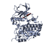
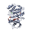
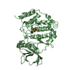
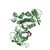
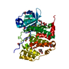
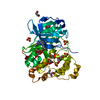
| Structure viewer | Molecule: MolmilJmol/JSmol |
|---|
- Downloads & links
-Download
| PDBx/mmCIF format | 5m4u.cif.gz | 164.4 KB | Display | PDBx/mmCIF format |
|---|---|---|---|---|
| PDB format | pdb5m4u.ent.gz | 126.6 KB | Display | PDB format |
| PDBx/mmJSON format | 5m4u.json.gz | Tree view | PDBx/mmJSON format | |
| Others |  Other downloads Other downloads |
-Validation report
| Arichive directory | https://data.pdbj.org/pub/pdb/validation_reports/m4/5m4uftp://data.pdbj.org/pub/pdb/validation_reports/m4/5m4u | HTTPS FTP |
|---|
-Related structure data
| Related structure data | 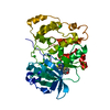 5m44C  5m4cC  5m4fC  5m4iC  5m56C  3ofmS S: Starting model for refinement C: citing same article ( |
|---|---|
| Similar structure data |


-Links
 PDBj
PDBj


- Assembly
| Deposited unit | 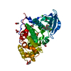
| ||||||||
|---|---|---|---|---|---|---|---|---|---|
| 1 |
| ||||||||
| Unit cell |
|
-Components
-Protein , 1 types, 1 molecules A
| #1: Protein | Mass: 42821.832 Da / Num. of mol.: 1 / Mutation: Asp39Gly, Cys336Ser Source method: isolated from a genetically manipulated source Source: (gene. exp.) Homo sapiens (human) / Gene: CSNK2A2, CK2A2 / Production host:  References: UniProt: P19784, non-specific serine/threonine protein kinase |
|---|
-Non-polymers , 6 types, 326 molecules 


| #2: Chemical | ChemComp-7FC /  Mass: 351.138 Da / Num. of mol.: 1 / Source method: obtained synthetically / Formula: C16H8Cl2O5 Mass: 351.138 Da / Num. of mol.: 1 / Source method: obtained synthetically / Formula: C16H8Cl2O5 | ||||||||
|---|---|---|---|---|---|---|---|---|---|
| #3: Chemical | ChemComp-GOL /  Mass: 92.094 Da / Num. of mol.: 5 / Source method: obtained synthetically / Formula: C3H8O3 Mass: 92.094 Da / Num. of mol.: 5 / Source method: obtained synthetically / Formula: C3H8O3#4: Chemical |  Mass: 59.044 Da / Num. of mol.: 3 / Source method: obtained synthetically / Formula: C2H3O2 Mass: 59.044 Da / Num. of mol.: 3 / Source method: obtained synthetically / Formula: C2H3O2#5: Chemical | ChemComp-CL / |  Mass: 35.453 Da / Num. of mol.: 1 / Source method: obtained synthetically / Formula: Cl Mass: 35.453 Da / Num. of mol.: 1 / Source method: obtained synthetically / Formula: Cl#6: Chemical | ChemComp-SO4 / |  Mass: 96.063 Da / Num. of mol.: 1 / Source method: obtained synthetically / Formula: SO4 Mass: 96.063 Da / Num. of mol.: 1 / Source method: obtained synthetically / Formula: SO4#7: Water | ChemComp-HOH / | Mass: 18.015 Da / Num. of mol.: 315 / Source method: isolated from a natural source / Formula: H2O |
-Experimental details
-Experiment
| Experiment | Method: X-RAY DIFFRACTION / Number of used crystals: 1 |
|---|
- Sample preparation
| Crystal | Density Matthews: 3.32 Å3/Da / Density % sol: 62.99 % |
|---|---|
| Crystal grow | Temperature: 277 K / Method: vapor diffusion, sitting drop / pH: 6.5 Details: PROTEIN STOCK SOLUTION: 5.5 MG/ML CK2ALPHA'D39G-C336S IN 0.5 M NACL, 25 MM TRIS/HCL, PH 8.5; INHIBITOR STOCK SOLUTION: 10 MM INHIBITOR IN DMSO; PROTEIN/INHIBITOR COMPLEX SOLUTION: 90 ...Details: PROTEIN STOCK SOLUTION: 5.5 MG/ML CK2ALPHA'D39G-C336S IN 0.5 M NACL, 25 MM TRIS/HCL, PH 8.5; INHIBITOR STOCK SOLUTION: 10 MM INHIBITOR IN DMSO; PROTEIN/INHIBITOR COMPLEX SOLUTION: 90 MICROLITER PROTEIN STOCK SOLUTION + 10 MICROLITER INHIBITOR STOCK SOLUTION; RESERVOIR SOLUTION: 25 % PEG5000 MME, 0.2 M ammonium sulphate, 0.1 M MES buffer, pH 6.5; DROP SOLUTION BEFORE EQULIBRATION: 0.3 MICROLITER PROTEIN/INHIBITOR COMPLEX SOLUTION + 0.3 MICROLITER RESERVOIR SOLUTION |
-Data collection
| Diffraction | Mean temperature: 100 K |
|---|---|
| Diffraction source | Source: SYNCHROTRON / Site: SLS  / Beamline: X06DA / Wavelength: 1 Å / Beamline: X06DA / Wavelength: 1 Å |
| Detector | Type: DECTRIS PILATUS 2M / Detector: PIXEL / Date: Oct 20, 2012 |
| Radiation | Protocol: SINGLE WAVELENGTH / Monochromatic (M) / Laue (L): M / Scattering type: x-ray |
| Radiation wavelength | Wavelength: 1 Å / Relative weight: 1 |
| Reflection | Resolution: 2.195→44.6 Å / Num. obs: 29246 / % possible obs: 99 % / Redundancy: 6.4 % / Biso Wilson estimate: 21.44 Å2 / CC1/2: 0.998 / Rmerge(I) obs: 0.1909 / Rsym value: 0.1909 / Net I/σ(I): 9.99 |
| Reflection shell | Resolution: 2.195→2.274 Å / Redundancy: 5.6 % / Rmerge(I) obs: 1.185 / Mean I/σ(I) obs: 1.72 / CC1/2: 0.684 / % possible all: 93 |
- Processing
| Software |
| |||||||||||||||||||||||||||||||||||||||||||||||||||||||||||||||||||||||||||
|---|---|---|---|---|---|---|---|---|---|---|---|---|---|---|---|---|---|---|---|---|---|---|---|---|---|---|---|---|---|---|---|---|---|---|---|---|---|---|---|---|---|---|---|---|---|---|---|---|---|---|---|---|---|---|---|---|---|---|---|---|---|---|---|---|---|---|---|---|---|---|---|---|---|---|---|---|
| Refinement | Method to determine structure: MOLECULAR REPLACEMENT Starting model: 3OFM Resolution: 2.195→44.502 Å / SU ML: 0.23 / Cross valid method: FREE R-VALUE / σ(F): 1.35 / Phase error: 21.03
| |||||||||||||||||||||||||||||||||||||||||||||||||||||||||||||||||||||||||||
| Solvent computation | Shrinkage radii: 0.9 Å / VDW probe radii: 1.11 Å | |||||||||||||||||||||||||||||||||||||||||||||||||||||||||||||||||||||||||||
| Refinement step | Cycle: LAST / Resolution: 2.195→44.502 Å
| |||||||||||||||||||||||||||||||||||||||||||||||||||||||||||||||||||||||||||
| Refine LS restraints |
| |||||||||||||||||||||||||||||||||||||||||||||||||||||||||||||||||||||||||||
| LS refinement shell |
| |||||||||||||||||||||||||||||||||||||||||||||||||||||||||||||||||||||||||||
| Refinement TLS params. | Method: refined / Refine-ID: X-RAY DIFFRACTION
| |||||||||||||||||||||||||||||||||||||||||||||||||||||||||||||||||||||||||||
| Refinement TLS group |
|
