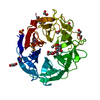
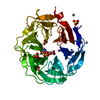
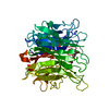
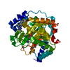
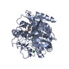
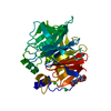
Entry Database : PDB / ID : 5m1zTitle STRUCTURE OF THE ALPHA-L-ARABINOFURANOSIDASE ARB93A FROM FUSARIUM GRAMINEARUM IN COMPLEX WITH AN hydroximolactone INHIBITOR Exo-1,5-alpha-L-arabinofuranobiosidase Keywords / / / Function / homology / / / / / / / / Biological species Gibberella zeae (fungus)Method / / / Resolution : 2 Å Authors Varrot, A. Funding support Organization Grant number Country National Health and Medical Research Council APP1073250
Journal : Chembiochem / Year : 2017Title : Exploiting sp(2) -Hybridisation in the Development of Potent 1,5-alpha-l-Arabinanase Inhibitors.Authors : Coyle, T. / Debowski, A.W. / Varrot, A. / Stubbs, K.A. History Deposition Oct 11, 2016 Deposition site / Processing site Revision 1.0 Dec 28, 2016 Provider / Type Revision 1.1 Mar 22, 2017 Group Revision 1.2 May 3, 2017 Group Revision 1.3 Jun 14, 2017 Group / Category Item _citation.country / _citation.journal_volume ... _citation.country / _citation.journal_volume / _citation.page_first / _citation.page_last Revision 2.0 Jul 29, 2020 Group Atomic model / Data collection ... Atomic model / Data collection / Derived calculations / Structure summary Category atom_site / chem_comp ... atom_site / chem_comp / entity / pdbx_branch_scheme / pdbx_chem_comp_identifier / pdbx_entity_branch / pdbx_entity_branch_descriptor / pdbx_entity_branch_link / pdbx_entity_branch_list / pdbx_entity_nonpoly / pdbx_nonpoly_scheme / pdbx_struct_assembly_gen / pdbx_struct_conn_angle / pdbx_struct_special_symmetry / struct_asym / struct_conn / struct_conn_type / struct_site / struct_site_gen Item _atom_site.auth_asym_id / _atom_site.auth_atom_id ... _atom_site.auth_asym_id / _atom_site.auth_atom_id / _atom_site.auth_seq_id / _atom_site.label_asym_id / _atom_site.label_atom_id / _atom_site.label_entity_id / _chem_comp.name / _chem_comp.type / _pdbx_struct_assembly_gen.asym_id_list / _pdbx_struct_conn_angle.ptnr1_auth_seq_id / _pdbx_struct_conn_angle.ptnr1_label_asym_id / _pdbx_struct_conn_angle.ptnr2_label_asym_id / _pdbx_struct_conn_angle.ptnr3_auth_seq_id / _pdbx_struct_conn_angle.ptnr3_label_asym_id / _pdbx_struct_conn_angle.value / _pdbx_struct_special_symmetry.label_asym_id / _struct_conn.conn_type_id / _struct_conn.id / _struct_conn.pdbx_dist_value / _struct_conn.pdbx_leaving_atom_flag / _struct_conn.ptnr1_auth_asym_id / _struct_conn.ptnr1_auth_comp_id / _struct_conn.ptnr1_auth_seq_id / _struct_conn.ptnr1_label_asym_id / _struct_conn.ptnr1_label_atom_id / _struct_conn.ptnr1_label_comp_id / _struct_conn.ptnr1_label_seq_id / _struct_conn.ptnr2_auth_asym_id / _struct_conn.ptnr2_auth_comp_id / _struct_conn.ptnr2_auth_seq_id / _struct_conn.ptnr2_label_asym_id / _struct_conn.ptnr2_label_atom_id / _struct_conn.ptnr2_label_comp_id / _struct_conn.ptnr2_symmetry / _struct_conn_type.id Description / Provider / Type Revision 2.1 Jan 17, 2024 Group Data collection / Database references ... Data collection / Database references / Derived calculations / Refinement description / Structure summary Category chem_comp / chem_comp_atom ... chem_comp / chem_comp_atom / chem_comp_bond / database_2 / pdbx_initial_refinement_model / struct_conn Item _chem_comp.pdbx_synonyms / _database_2.pdbx_DOI ... _chem_comp.pdbx_synonyms / _database_2.pdbx_DOI / _database_2.pdbx_database_accession / _struct_conn.pdbx_leaving_atom_flag
Show all Show less
 Movie
Movie Controller

 Yorodumi
 Open data
 Basic information
 Components
 Keywords
 Function and homology information
 Gibberella zeae (fungus)
 X-RAY DIFFRACTION /
  Authors
 Australia, 1items
  Citation
 Structure visualization
 Downloads & links
 Other downloads
 PDBj
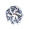
PDBj Assembly
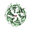
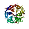


 Mass: 96.063 Da / Num. of mol.: 5 / Source method: obtained synthetically / Formula: SO4
 Mass: 54.938 Da / Num. of mol.: 4 / Source method: obtained synthetically / Formula: Mn
 Mass: 62.068 Da / Num. of mol.: 1 / Source method: obtained synthetically / Formula: C2H6O2
 Mass: 92.094 Da / Num. of mol.: 1 / Source method: obtained synthetically / Formula: C3H8O3
 Sample preparation
 / Beamline: ID23-1 / Wavelength: 1.12713 Å
 Processing