 Movie
Movie Controller

[English] 日本語
 Yorodumi
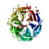
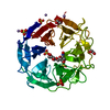
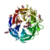
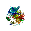
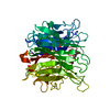
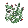
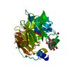
- PDB-2ydp: Structure of the E242A mutant of the alpha-l-arabinofuranosidase ... -
+ Open data

- Basic information
| Entry | Database: PDB / ID: 2ydp | |||||||||
|---|---|---|---|---|---|---|---|---|---|---|
| Title | Structure of the E242A mutant of the alpha-l-arabinofuranosidase arb93a from fusarium graminearum in complex with an iminosugar inhibitor | |||||||||
 Components Components | EXO-1,5-ALPHA-L-ARABINOFURANOBIOSIDASE | |||||||||
 Keywords Keywords | HYDROLASE / GLYCOSYL HYDROLASE | |||||||||
| Function / homology |  Function and homology information Function and homology informationnon-reducing end alpha-L-arabinofuranosidase / alpha-L-arabinofuranosidase activity / metal ion binding Similarity search - Function | |||||||||
| Biological species |  GIBBERELLA ZEAE (fungus) GIBBERELLA ZEAE (fungus) | |||||||||
| Method |  X-RAY DIFFRACTION / SYNCHROTRON / MOLECULAR REPLACEMENT / Resolution: 1.85 Å X-RAY DIFFRACTION / SYNCHROTRON / MOLECULAR REPLACEMENT / Resolution: 1.85 Å | |||||||||
 Authors Authors | Goddard-Borger, E.D. / Carapito, R. / Jeltsch, J.M. / Phalip, V. / Stick, R.V. / Varrot, A. | |||||||||
 Citation Citation | Journal: Chem.Commun.(Camb.) / Year: 2011 Title: Alpha-L-Arabinofuranosylated Pyrrolidines as Arabinanase Inhibitors. Authors: Goddard-Borger, E.D. / Carapito, R. / Jeltsch, J. / Phalip, V. / Stick, R.V. / Varrot, A. | |||||||||
| History |
|
- Structure visualization
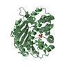
| Structure viewer | Molecule: MolmilJmol/JSmol |
|---|
- Downloads & links
-Download
| PDBx/mmCIF format | 2ydp.cif.gz | 243.9 KB | Display | PDBx/mmCIF format |
|---|---|---|---|---|
| PDB format | pdb2ydp.ent.gz | 193.6 KB | Display | PDB format |
| PDBx/mmJSON format | 2ydp.json.gz | Tree view | PDBx/mmJSON format | |
| Others |  Other downloads Other downloads |
-Validation report
| Arichive directory | https://data.pdbj.org/pub/pdb/validation_reports/yd/2ydpftp://data.pdbj.org/pub/pdb/validation_reports/yd/2ydp | HTTPS FTP |
|---|
-Related structure data
| Related structure data |  2ydtC 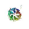 2w5nS C: citing same article ( S: Starting model for refinement |
|---|---|
| Similar structure data |

-Links
 PDBj
PDBj- Assembly
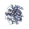
| Deposited unit | 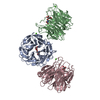
| ||||||||
|---|---|---|---|---|---|---|---|---|---|
| 1 | 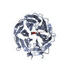
| ||||||||
| 2 | 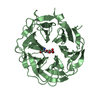
| ||||||||
| 3 | 
| ||||||||
| Unit cell |
|
-Components
| #1: Protein | Mass: 41063.324 Da / Num. of mol.: 3 / Mutation: YES Source method: isolated from a genetically manipulated source Source: (gene. exp.) GIBBERELLA ZEAE (fungus) / Production host:  References: UniProt: B8ZY56, non-reducing end alpha-L-arabinofuranosidase #2: Chemical | 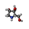  Mass: 133.146 Da / Num. of mol.: 3 / Source method: obtained synthetically / Formula: C5H11NO3 Mass: 133.146 Da / Num. of mol.: 3 / Source method: obtained synthetically / Formula: C5H11NO3#3: Sugar | 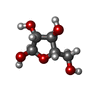  Type: L-saccharide, alpha linking / Mass: 150.130 Da / Num. of mol.: 3 Type: L-saccharide, alpha linking / Mass: 150.130 Da / Num. of mol.: 3Source method: isolated from a genetically manipulated source Formula: C5H10O5 #4: Chemical |   Mass: 40.078 Da / Num. of mol.: 3 / Source method: obtained synthetically / Formula: Ca Mass: 40.078 Da / Num. of mol.: 3 / Source method: obtained synthetically / Formula: Ca#5: Water | ChemComp-HOH / |  Mass: 18.015 Da / Num. of mol.: 964 / Source method: isolated from a natural source / Formula: H2O Mass: 18.015 Da / Num. of mol.: 964 / Source method: isolated from a natural source / Formula: H2OCompound details | ENGINEERED RESIDUE IN CHAIN A, GLU 226 TO ALA ENGINEERED RESIDUE IN CHAIN B, GLU 226 TO ALA ...ENGINEERED | Sequence details | THE SEQUENCE CORRESPOND | |
|---|
-Experimental details
-Experiment
| Experiment | Method: X-RAY DIFFRACTION / Number of used crystals: 1 |
|---|
- Sample preparation
| Crystal | Density Matthews: 2.22 Å3/Da / Density % sol: 44.64 % / Description: NONE |
|---|---|
| Crystal grow | pH: 6.5 Details: 20% PEG 8000, 100 MM SODIUM CACODYLATE PH 6.5, 200 MM LISO4, 20% GLYCEROL AS CYOPROTECTANT. |
-Data collection
| Diffraction | Mean temperature: 100 K |
|---|---|
| Diffraction source | Source: SYNCHROTRON / Site: ESRF  / Beamline: ID14-4 / Wavelength: 0.981 / Beamline: ID14-4 / Wavelength: 0.981 |
| Detector | Type: ADSC CCD / Detector: CCD / Date: Jun 19, 2009 |
| Radiation | Protocol: SINGLE WAVELENGTH / Monochromatic (M) / Laue (L): M / Scattering type: x-ray |
| Radiation wavelength | Wavelength: 0.981 Å / Relative weight: 1 |
| Reflection | Resolution: 1.85→35.47 Å / Num. obs: 86184 / % possible obs: 95.2 % / Observed criterion σ(I): 1.8 / Redundancy: 2 % / Rmerge(I) obs: 0.1 / Net I/σ(I): 5.5 |
| Reflection shell | Resolution: 1.85→1.95 Å / Redundancy: 2 % / Rmerge(I) obs: 0.37 / Mean I/σ(I) obs: 1.8 / % possible all: 95.2 |
- Processing
| Software |
| ||||||||||||||||||||||||||||||||||||||||||||||||||||||||||||||||||||||||||||||||||||||||||||||||||||||||||||||||||||||||||||||||||||||||||||||||||||||||||||||||||||||||||||||||||||||
|---|---|---|---|---|---|---|---|---|---|---|---|---|---|---|---|---|---|---|---|---|---|---|---|---|---|---|---|---|---|---|---|---|---|---|---|---|---|---|---|---|---|---|---|---|---|---|---|---|---|---|---|---|---|---|---|---|---|---|---|---|---|---|---|---|---|---|---|---|---|---|---|---|---|---|---|---|---|---|---|---|---|---|---|---|---|---|---|---|---|---|---|---|---|---|---|---|---|---|---|---|---|---|---|---|---|---|---|---|---|---|---|---|---|---|---|---|---|---|---|---|---|---|---|---|---|---|---|---|---|---|---|---|---|---|---|---|---|---|---|---|---|---|---|---|---|---|---|---|---|---|---|---|---|---|---|---|---|---|---|---|---|---|---|---|---|---|---|---|---|---|---|---|---|---|---|---|---|---|---|---|---|---|---|
| Refinement | Method to determine structure: MOLECULAR REPLACEMENT Starting model: PDB ENTRY 2W5N Resolution: 1.85→33.97 Å / Cor.coef. Fo:Fc: 0.963 / Cor.coef. Fo:Fc free: 0.937 / SU B: 3.631 / SU ML: 0.107 / Cross valid method: THROUGHOUT / ESU R: 0.154 / ESU R Free: 0.148 / Stereochemistry target values: MAXIMUM LIKELIHOOD / Details: HYDROGENS HAVE BEEN ADDED IN THE RIDING POSITIONS
| ||||||||||||||||||||||||||||||||||||||||||||||||||||||||||||||||||||||||||||||||||||||||||||||||||||||||||||||||||||||||||||||||||||||||||||||||||||||||||||||||||||||||||||||||||||||
| Solvent computation | Ion probe radii: 0.8 Å / Shrinkage radii: 0.8 Å / VDW probe radii: 1.4 Å / Solvent model: MASK | ||||||||||||||||||||||||||||||||||||||||||||||||||||||||||||||||||||||||||||||||||||||||||||||||||||||||||||||||||||||||||||||||||||||||||||||||||||||||||||||||||||||||||||||||||||||
| Displacement parameters | Biso mean: 21.56 Å2
| ||||||||||||||||||||||||||||||||||||||||||||||||||||||||||||||||||||||||||||||||||||||||||||||||||||||||||||||||||||||||||||||||||||||||||||||||||||||||||||||||||||||||||||||||||||||
| Refinement step | Cycle: LAST / Resolution: 1.85→33.97 Å
| ||||||||||||||||||||||||||||||||||||||||||||||||||||||||||||||||||||||||||||||||||||||||||||||||||||||||||||||||||||||||||||||||||||||||||||||||||||||||||||||||||||||||||||||||||||||
| Refine LS restraints |
|