 Movie
Movie Controller

[English] 日本語
 Yorodumi
- PDB-5m0j: Crystal structure of the cytoplasmic complex with She2p, She3p, a... -
+ Open data

- Basic information
| Entry | Database: PDB / ID: 5m0j | ||||||
|---|---|---|---|---|---|---|---|
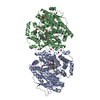
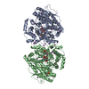
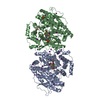
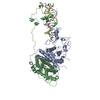
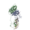
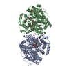
| Title | Crystal structure of the cytoplasmic complex with She2p, She3p, and the ASH1 mRNA E3-localization element | ||||||
 Components Components |
| ||||||
 Keywords Keywords | RNA BINDING PROTEIN / She2p / She3p / ASH1-mRNA / mRNA transport | ||||||
| Function / homology |  Function and homology information Function and homology informationmating type switching / endoplasmic reticulum inheritance / cellular bud tip / intracellular mRNA localization / sequence-specific mRNA binding / mRNA transport / mRNA binding / lipid binding / endoplasmic reticulum membrane / RNA binding ...mating type switching / endoplasmic reticulum inheritance / cellular bud tip / intracellular mRNA localization / sequence-specific mRNA binding / mRNA transport / mRNA binding / lipid binding / endoplasmic reticulum membrane / RNA binding / nucleus / cytosol / cytoplasm Similarity search - Function | ||||||
| Biological species |  | ||||||
| Method |  X-RAY DIFFRACTION / SYNCHROTRON / MOLECULAR REPLACEMENT / Resolution: 2.8 Å X-RAY DIFFRACTION / SYNCHROTRON / MOLECULAR REPLACEMENT / Resolution: 2.8 Å | ||||||
 Authors Authors | Edelmann, F.T. / Janowski, R. / Niessing, D. | ||||||
 Citation Citation | Journal: Nat. Struct. Mol. Biol. / Year: 2017 Title: Molecular architecture and dynamics of ASH1 mRNA recognition by its mRNA-transport complex. Authors: Edelmann, F.T. / Schlundt, A. / Heym, R.G. / Jenner, A. / Niedner-Boblenz, A. / Syed, M.I. / Paillart, J.C. / Stehle, R. / Janowski, R. / Sattler, M. / Jansen, R.P. / Niessing, D. | ||||||
| History |
|
- Structure visualization
| Structure viewer | Molecule: MolmilJmol/JSmol |
|---|
- Downloads & links
-Download
| PDBx/mmCIF format | 5m0j.cif.gz | 269.4 KB | Display | PDBx/mmCIF format |
|---|---|---|---|---|
| PDB format | pdb5m0j.ent.gz | 204.1 KB | Display | PDB format |
| PDBx/mmJSON format | 5m0j.json.gz | Tree view | PDBx/mmJSON format | |
| Others |  Other downloads Other downloads |
-Validation report
| Arichive directory | https://data.pdbj.org/pub/pdb/validation_reports/m0/5m0jftp://data.pdbj.org/pub/pdb/validation_reports/m0/5m0j | HTTPS FTP |
|---|
-Related structure data
| Related structure data | 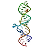 5m0hC 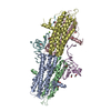 5m0iC  1xlyS C: citing same article ( S: Starting model for refinement |
|---|---|
| Similar structure data |


-Links
 PDBj
PDBj


- Assembly
| Deposited unit | 
| |||||||||||||||||||||||||||||||||||||||||||||||||||||||||||||||||||||||||||||||||||||||||||||||||||||||||||||||||||||||||||||||||||||||||||||||||||||||||||||||||||||||||||||||||||||||||||||
|---|---|---|---|---|---|---|---|---|---|---|---|---|---|---|---|---|---|---|---|---|---|---|---|---|---|---|---|---|---|---|---|---|---|---|---|---|---|---|---|---|---|---|---|---|---|---|---|---|---|---|---|---|---|---|---|---|---|---|---|---|---|---|---|---|---|---|---|---|---|---|---|---|---|---|---|---|---|---|---|---|---|---|---|---|---|---|---|---|---|---|---|---|---|---|---|---|---|---|---|---|---|---|---|---|---|---|---|---|---|---|---|---|---|---|---|---|---|---|---|---|---|---|---|---|---|---|---|---|---|---|---|---|---|---|---|---|---|---|---|---|---|---|---|---|---|---|---|---|---|---|---|---|---|---|---|---|---|---|---|---|---|---|---|---|---|---|---|---|---|---|---|---|---|---|---|---|---|---|---|---|---|---|---|---|---|---|---|---|---|---|
| 1 |
| |||||||||||||||||||||||||||||||||||||||||||||||||||||||||||||||||||||||||||||||||||||||||||||||||||||||||||||||||||||||||||||||||||||||||||||||||||||||||||||||||||||||||||||||||||||||||||||
| Unit cell |
| |||||||||||||||||||||||||||||||||||||||||||||||||||||||||||||||||||||||||||||||||||||||||||||||||||||||||||||||||||||||||||||||||||||||||||||||||||||||||||||||||||||||||||||||||||||||||||||
| Noncrystallographic symmetry (NCS) | NCS domain:
NCS domain segments: Component-ID: _ / Refine code: _
|