 Movie
Movie Controller


+ Open data

- Basic information
| Entry | Database: PDB / ID: 1xly | ||||||
|---|---|---|---|---|---|---|---|
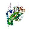
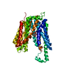
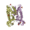
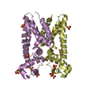
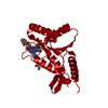
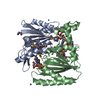
| Title | X-RAY STRUCTURE OF THE RNA-BINDING PROTEIN SHE2p | ||||||
 Components Components | SHE2p | ||||||
 Keywords Keywords | RNA BINDING PROTEIN / basic helical hairpin / five helix bundle / dimer / RNA-BINDING PROTEIN | ||||||
| Function / homology |  Function and homology information Function and homology informationmating type switching / cellular bud tip / intracellular mRNA localization / sequence-specific mRNA binding / mRNA transport / mRNA binding / lipid binding / nucleus / cytoplasm / cytosol Similarity search - Function | ||||||
| Biological species |  | ||||||
| Method |  X-RAY DIFFRACTION / SYNCHROTRON / MIR / Resolution: 1.95 Å X-RAY DIFFRACTION / SYNCHROTRON / MIR / Resolution: 1.95 Å | ||||||
 Authors Authors | Niessing, D. / Huettelmaier, S. / Zenklusen, D. / Singer, R.H. / Burley, S.K. | ||||||
 Citation Citation | Journal: Cell(Cambridge,Mass.) / Year: 2004 Title: She2p is a novel RNA binding protein with a basic helical hairpin motif Authors: Niessing, D. / Huettelmaier, S. / Zenklusen, D. / Singer, R.H. / Burley, S.K. | ||||||
| History |
|
- Structure visualization
| Structure viewer | Molecule: MolmilJmol/JSmol |
|---|
- Downloads & links
-Download
| PDBx/mmCIF format | 1xly.cif.gz | 101.1 KB | Display | PDBx/mmCIF format |
|---|---|---|---|---|
| PDB format | pdb1xly.ent.gz | 78.3 KB | Display | PDB format |
| PDBx/mmJSON format | 1xly.json.gz | Tree view | PDBx/mmJSON format | |
| Others |  Other downloads Other downloads |
-Validation report
| Arichive directory | https://data.pdbj.org/pub/pdb/validation_reports/xl/1xlyftp://data.pdbj.org/pub/pdb/validation_reports/xl/1xly | HTTPS FTP |
|---|
-Related structure data
| Similar structure data |
|---|


-Links
 PDBj
PDBj- Assembly
| Deposited unit | 
| ||||||||
|---|---|---|---|---|---|---|---|---|---|
| 1 |
| ||||||||
| Unit cell |
| ||||||||
| Details | The biological assembly is a dimer of non-crystallographic symmetry |
-Components
| #1: Protein | Mass: 26807.252 Da / Num. of mol.: 2 / Mutation: C14S, C68, C106S, C180S Source method: isolated from a genetically manipulated source Source: (gene. exp.) Gene: SHE2, YKL130C / Plasmid: pGEX-6P1 / Production host:  #2: Water | ChemComp-HOH / |  Mass: 18.015 Da / Num. of mol.: 302 / Source method: isolated from a natural source / Formula: H2O Mass: 18.015 Da / Num. of mol.: 302 / Source method: isolated from a natural source / Formula: H2O |
|---|
-Experimental details
-Experiment
| Experiment | Method: X-RAY DIFFRACTION / Number of used crystals: 1 |
|---|
- Sample preparation
| Crystal | Density Matthews: 2.41 Å3/Da / Density % sol: 49 % |
|---|---|
| Crystal grow | Temperature: 293 K / pH: 8.5 Details: PEG 4000, lithium sulfate, tris, beta-octyl-glucoside, pH 8.5, VAPOR DIFFUSION, HANGING DROP, temperature 293K |
-Data collection
| Diffraction | Mean temperature: 100 K |
|---|---|
| Diffraction source | Source: SYNCHROTRON / Site: APS  / Beamline: 31-ID / Wavelength: 1.072 / Beamline: 31-ID / Wavelength: 1.072 |
| Detector | Type: MARRESEARCH / Detector: CCD / Date: Oct 23, 2003 |
| Radiation | Monochromator: DIAMOND 111 / Protocol: SINGLE WAVELENGTH / Monochromatic (M) / Laue (L): M / Scattering type: x-ray |
| Radiation wavelength | Wavelength: 1.072 Å / Relative weight: 1 |
| Reflection | Resolution: 1.95→25 Å / Num. obs: 37528 / % possible obs: 98 % / Observed criterion σ(I): 0 / Biso Wilson estimate: 24.1 Å2 / Rsym value: 0.086 / Net I/σ(I): 11.1 |
| Reflection shell | Resolution: 1.95→2.02 Å / Mean I/σ(I) obs: 2.7 / Rsym value: 0.486 / % possible all: 97.2 |
- Processing
| Software |
| ||||||||||||||||||||||||||||||||||||||||||||||||||||||||||||||||||||||||||||||||||||
|---|---|---|---|---|---|---|---|---|---|---|---|---|---|---|---|---|---|---|---|---|---|---|---|---|---|---|---|---|---|---|---|---|---|---|---|---|---|---|---|---|---|---|---|---|---|---|---|---|---|---|---|---|---|---|---|---|---|---|---|---|---|---|---|---|---|---|---|---|---|---|---|---|---|---|---|---|---|---|---|---|---|---|---|---|---|
| Refinement | Method to determine structure: MIR / Resolution: 1.95→19.76 Å / Cross valid method: THROUGHOUT / σ(F): 0 / Stereochemistry target values: Engh & Huber Details: PHE 176 in CHAIN B has very high B values and is close to the end of a short helix. It is well possible, that this residue adopts different conformations in the various molecules of the ...Details: PHE 176 in CHAIN B has very high B values and is close to the end of a short helix. It is well possible, that this residue adopts different conformations in the various molecules of the asymmetric, so that in reality the reported clashes are avoided. The high B values and the weak visible electron density prevent a clear observation in this regard. We, therefore, chose to refine a conformation that is most consistent with the visible density and conforms with stereochemical requirements. It is also worth mentioning that, for the second molecule of this homodimer (CHAIN A), this region shows much stronger electron density. The structural model of CHAIN A, therefore, gives a very clear, and much better, description of this region. Consistently, we show in our publication a symmetry operated homodimer consisting of two CHAIN A models.
| ||||||||||||||||||||||||||||||||||||||||||||||||||||||||||||||||||||||||||||||||||||
| Displacement parameters | Biso mean: 44.5 Å2 | ||||||||||||||||||||||||||||||||||||||||||||||||||||||||||||||||||||||||||||||||||||
| Refinement step | Cycle: LAST / Resolution: 1.95→19.76 Å
| ||||||||||||||||||||||||||||||||||||||||||||||||||||||||||||||||||||||||||||||||||||
| Refine LS restraints |
|
