 Movie
Movie Controller


+ Open data

- Basic information
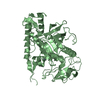
| Entry | Database: PDB / ID: 7kyf | ||||||
|---|---|---|---|---|---|---|---|
| Title | Botulism Neurooxin Light Chain A app form | ||||||
 Components Components | Bont/A1 | ||||||
 Keywords Keywords | TOXIN / BoNT / botulinum neurotoxin nva-ila dipetpide bound | ||||||
| Function / homology |  Function and homology information Function and homology informationhost cell presynaptic membrane / host cell cytoplasmic vesicle / host cell cytosol / transmembrane protein transporter activity / metalloendopeptidase activity / toxin activity / proteolysis / extracellular region / zinc ion binding Similarity search - Function | ||||||
| Biological species |   Clostridium botulinum (bacteria) Clostridium botulinum (bacteria) | ||||||
| Method |  X-RAY DIFFRACTION / SYNCHROTRON / MOLECULAR REPLACEMENT / Resolution: 2.33 Å X-RAY DIFFRACTION / SYNCHROTRON / MOLECULAR REPLACEMENT / Resolution: 2.33 Å | ||||||
 Authors Authors | Ortega, M.E. / Salzameda, N.T. | ||||||
 Citation Citation | Journal: Acs Med.Chem.Lett. / Year: 2021 Title: Discovery of Dipeptides as Potent Botulinum Neurotoxin A Light-Chain Inhibitors. Authors: Amezcua, M. / Cruz, R.S. / Ku, A. / Moran, W. / Ortega, M.E. / Salzameda, N.T. | ||||||
| History |
|
- Structure visualization
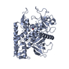
| Structure viewer | Molecule: MolmilJmol/JSmol |
|---|
- Downloads & links
-Download
| PDBx/mmCIF format | 7kyf.cif.gz | 96.9 KB | Display | PDBx/mmCIF format |
|---|---|---|---|---|
| PDB format | pdb7kyf.ent.gz | 71.3 KB | Display | PDB format |
| PDBx/mmJSON format | 7kyf.json.gz | Tree view | PDBx/mmJSON format | |
| Others |  Other downloads Other downloads |
-Validation report
| Arichive directory | https://data.pdbj.org/pub/pdb/validation_reports/ky/7kyfftp://data.pdbj.org/pub/pdb/validation_reports/ky/7kyf | HTTPS FTP |
|---|
-Related structure data
| Related structure data | 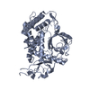 7ky2C 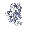 7kyhC 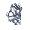 4hevS S: Starting model for refinement C: citing same article ( |
|---|---|
| Similar structure data |


-Links
 PDBj
PDBj- Assembly
| Deposited unit | 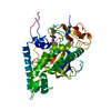
| ||||||||
|---|---|---|---|---|---|---|---|---|---|
| 1 |
| ||||||||
| Unit cell |
|
-Components
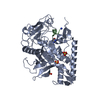
| #1: Protein | Mass: 47777.039 Da / Num. of mol.: 1 Source method: isolated from a genetically manipulated source Source: (gene. exp.) Clostridium botulinum (bacteria) / Production host: |
|---|---|
| #2: Chemical | ChemComp-ZN /   Mass: 65.409 Da / Num. of mol.: 1 / Source method: obtained synthetically / Formula: Zn Mass: 65.409 Da / Num. of mol.: 1 / Source method: obtained synthetically / Formula: Zn |
| #3: Chemical | ChemComp-XC1 /   Mass: 454.369 Da / Num. of mol.: 1 / Source method: obtained synthetically / Formula: C17H25Cl2N3O5S / Feature type: SUBJECT OF INVESTIGATION Mass: 454.369 Da / Num. of mol.: 1 / Source method: obtained synthetically / Formula: C17H25Cl2N3O5S / Feature type: SUBJECT OF INVESTIGATION |
| #4: Water | ChemComp-HOH /  Mass: 18.015 Da / Num. of mol.: 19 / Source method: isolated from a natural source / Formula: H2O Mass: 18.015 Da / Num. of mol.: 19 / Source method: isolated from a natural source / Formula: H2O |
| Has ligand of interest | Y |
-Experimental details
-Experiment
| Experiment | Method: X-RAY DIFFRACTION / Number of used crystals: 1 |
|---|
- Sample preparation
| Crystal | Density Matthews: 2.29 Å3/Da / Density % sol: 46.38 % |
|---|---|
| Crystal grow | Temperature: 298 K / Method: vapor diffusion, hanging drop Details: 50mM sodium cacodylate pH 7.0, 200mM ammonium sulfate, 30% PEG 8000 |
-Data collection
| Diffraction | Mean temperature: 100 K / Serial crystal experiment: N |
|---|---|
| Diffraction source | Source: SYNCHROTRON / Site: ALS  / Beamline: 4.2.2 / Wavelength: 0.97625 Å / Beamline: 4.2.2 / Wavelength: 0.97625 Å |
| Detector | Type: RDI CMOS_8M / Detector: CMOS / Date: Aug 20, 2020 |
| Radiation | Protocol: SINGLE WAVELENGTH / Monochromatic (M) / Laue (L): M / Scattering type: x-ray |
| Radiation wavelength | Wavelength: 0.97625 Å / Relative weight: 1 |
| Reflection | Resolution: 2→43.25 Å / Num. obs: 19587 / % possible obs: 93.2 % / Redundancy: 1 % / Rmerge(I) obs: 0.092 / Net I/σ(I): 12.5 |
- Processing
| Software |
| ||||||||||||||||||||||||||||||||||||||||||||||||||||||||||||
|---|---|---|---|---|---|---|---|---|---|---|---|---|---|---|---|---|---|---|---|---|---|---|---|---|---|---|---|---|---|---|---|---|---|---|---|---|---|---|---|---|---|---|---|---|---|---|---|---|---|---|---|---|---|---|---|---|---|---|---|---|---|
| Refinement | Method to determine structure: MOLECULAR REPLACEMENT Starting model: 4hev Resolution: 2.33→43.25 Å / Cor.coef. Fo:Fc: 0.958 / Cor.coef. Fo:Fc free: 0.945 / SU B: 2.217 / SU ML: 0.052 / Cross valid method: THROUGHOUT / σ(F): 0 / ESU R: 0.326 / ESU R Free: 0.186 / Stereochemistry target values: MAXIMUM LIKELIHOOD Details: HYDROGENS HAVE BEEN ADDED IN THE RIDING POSITIONS U VALUES : REFINED INDIVIDUALLY
| ||||||||||||||||||||||||||||||||||||||||||||||||||||||||||||
| Solvent computation | Ion probe radii: 0.8 Å / Shrinkage radii: 0.8 Å / VDW probe radii: 1.2 Å / Solvent model: MASK | ||||||||||||||||||||||||||||||||||||||||||||||||||||||||||||
| Displacement parameters | Biso max: 356.19 Å2 / Biso mean: 55.137 Å2 / Biso min: 8.92 Å2
| ||||||||||||||||||||||||||||||||||||||||||||||||||||||||||||
| Refinement step | Cycle: final / Resolution: 2.33→43.25 Å
| ||||||||||||||||||||||||||||||||||||||||||||||||||||||||||||
| Refine LS restraints |
| ||||||||||||||||||||||||||||||||||||||||||||||||||||||||||||
| LS refinement shell | Resolution: 2.33→2.391 Å / Rfactor Rfree error: 0 / Total num. of bins used: 20
|
