[English] 日本語
Yorodumi
- PDB-5lrv: Structure of Cezanne/OTUD7B OTU domain bound to Lys11-linked diub... -

+
Open data


ID or keywords:

Loading...

-
Basic information

Entry
Database: PDB / ID: 5lrv
Title
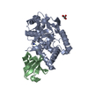
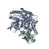
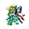
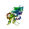
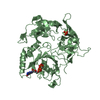
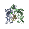
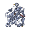
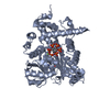
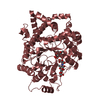
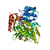
Structure of Cezanne/OTUD7B OTU domain bound to Lys11-linked diubiquitin
Components
  • (Polyubiquitin- ...) x 2
  • OTU domain-containing protein 7B
KeywordsHYDROLASE / protease / deubiquitinase / OTU domain
Function / homology
Function and homology information


protein deubiquitination involved in ubiquitin-dependent protein catabolic process / mucosal immune response / protein K11-linked deubiquitination / positive regulation of TORC2 signaling / negative regulation of interleukin-8 production / protein K48-linked deubiquitination / negative regulation of protein localization to nucleus / K48-linked deubiquitinase activity / protein K63-linked deubiquitination / symbiont entry into host cell via disruption of host cell glycocalyx ...protein deubiquitination involved in ubiquitin-dependent protein catabolic process / mucosal immune response / protein K11-linked deubiquitination / positive regulation of TORC2 signaling / negative regulation of interleukin-8 production / protein K48-linked deubiquitination / negative regulation of protein localization to nucleus / K48-linked deubiquitinase activity / protein K63-linked deubiquitination / symbiont entry into host cell via disruption of host cell glycocalyx / TNFR1-induced proapoptotic signaling / K63-linked polyubiquitin modification-dependent protein binding / symbiont entry into host cell via disruption of host cell envelope / virus tail / protein deubiquitination / negative regulation of TORC1 signaling / negative regulation of canonical NF-kappaB signal transduction / TNFR1-induced NF-kappa-B signaling pathway / cysteine-type peptidase activity / Regulation of TNFR1 signaling / Ovarian tumor domain proteases / adaptive immune response / in utero embryonic development / ubiquitinyl hydrolase 1 / cysteine-type deubiquitinase activity / negative regulation of transcription by RNA polymerase II / DNA binding / zinc ion binding / nucleus / cytosol / cytoplasm
Similarity search - Function
: / Zinc finger, A20-type / : / A20-like zinc finger / UBA-like domain / Zinc finger A20-type profile. / A20-like zinc fingers / OTU-like cysteine protease / OTU domain / OTU domain profile. ...: / Zinc finger, A20-type / : / A20-like zinc finger / UBA-like domain / Zinc finger A20-type profile. / A20-like zinc fingers / OTU-like cysteine protease / OTU domain / OTU domain profile. / Pectate lyase superfamily protein / Rhamnogalacturonase A/epimerase, pectate lyase-like / Pectin lyase fold / Pectin lyase fold/virulence factor / Ubiquitin family
Similarity search - Domain/homology
PHOSPHATE ION / Tail fiber / OTU domain-containing protein 7B
Similarity search - Component
Biological speciesHomo sapiens (human)
MethodX-RAY DIFFRACTION / SYNCHROTRON / MOLECULAR REPLACEMENT / Resolution: 2.8 Å
AuthorsMevissen, T.E.T. / Kulathu, Y. / Mulder, M.P.C. / Geurink, P.P. / Maslen, S.L. / Gersch, M. / Elliott, P.R. / Burke, J.E. / van Tol, B.D.M. / Akutsu, M. ...Mevissen, T.E.T. / Kulathu, Y. / Mulder, M.P.C. / Geurink, P.P. / Maslen, S.L. / Gersch, M. / Elliott, P.R. / Burke, J.E. / van Tol, B.D.M. / Akutsu, M. / El Oualid, F. / Kawasaki, M. / Freund, S.M.V. / Ovaa, H. / Komander, D.
Funding support United Kingdom, 2items
OrganizationGrant numberCountry
Medical Research Council (United Kingdom)U105192732 United Kingdom
European Research Council309756 United Kingdom
CitationJournal: Nature / Year: 2016
Title: Molecular basis of Lys11-polyubiquitin specificity in the deubiquitinase Cezanne.
Authors: Mevissen, T.E. / Kulathu, Y. / Mulder, M.P. / Geurink, P.P. / Maslen, S.L. / Gersch, M. / Elliott, P.R. / Burke, J.E. / van Tol, B.D. / Akutsu, M. / El Oualid, F. / Kawasaki, M. / Freund, S. ...Authors: Mevissen, T.E. / Kulathu, Y. / Mulder, M.P. / Geurink, P.P. / Maslen, S.L. / Gersch, M. / Elliott, P.R. / Burke, J.E. / van Tol, B.D. / Akutsu, M. / El Oualid, F. / Kawasaki, M. / Freund, S.M. / Ovaa, H. / Komander, D.
History
DepositionAug 22, 2016Deposition site: PDBE / Processing site: PDBE
Revision 1.0Oct 19, 2016Provider: repository / Type: Initial release
Revision 1.1Oct 26, 2016Group: Database references
Revision 1.2Sep 13, 2017Group: Author supporting evidence / Category: pdbx_audit_support / Item: _pdbx_audit_support.funding_organization
Revision 2.0Nov 15, 2023Group: Atomic model / Data collection ...Atomic model / Data collection / Database references / Derived calculations
Category: atom_site / atom_site_anisotrop ...atom_site / atom_site_anisotrop / chem_comp_atom / chem_comp_bond / database_2 / struct_conn
Item: _atom_site.auth_atom_id / _atom_site.label_atom_id ..._atom_site.auth_atom_id / _atom_site.label_atom_id / _atom_site_anisotrop.pdbx_auth_atom_id / _atom_site_anisotrop.pdbx_label_atom_id / _database_2.pdbx_DOI / _database_2.pdbx_database_accession / _struct_conn.ptnr2_label_atom_id
Revision 2.1Nov 13, 2024Group: Structure summary / Category: pdbx_entry_details / pdbx_modification_feature

-
Structure visualization

Structure viewerMolecule:
MolmilJmol/JSmol

Downloads & links

-
Assembly

Deposited unit
A: OTU domain-containing protein 7B
B: Polyubiquitin-B
C: Polyubiquitin-B
hetero molecules


Theoretical massNumber of molelcules
Total (without water)53,9768
Polymers53,5133
Non-polymers4635
Water543
1


  • Idetical with deposited unit
  • defined by software
TypeNameSymmetry operationNumber
identity operation1_555x,y,z1
Buried area5110 Å2
ΔGint-28 kcal/mol
Surface area18470 Å2
MethodPISA
Unit cell
Length a, b, c (Å)92.030, 56.520, 91.750
Angle α, β, γ (deg.)90.00, 90.00, 90.00
Int Tables number19
Space group name H-MP212121

-
Components

-
Protein , 1 types, 1 molecules A

#1: Protein OTU domain-containing protein 7B / Cellular zinc finger anti-NF-kappa-B protein / Zinc finger A20 domain-containing protein 1 / Zinc ...Cellular zinc finger anti-NF-kappa-B protein / Zinc finger A20 domain-containing protein 1 / Zinc finger protein Cezanne


Mass: 36360.375 Da / Num. of mol.: 1
Source method: isolated from a genetically manipulated source
Source: (gene. exp.) Homo sapiens (human) / Gene: OTUD7B, ZA20D1 / Production host: Escherichia coli (E. coli) / References: UniProt: Q6GQQ9, ubiquitinyl hydrolase 1

-
Polyubiquitin- ... , 2 types, 2 molecules BC

#2: Protein Polyubiquitin-B


Mass: 8547.770 Da / Num. of mol.: 1 / Mutation: K11X / Source method: obtained synthetically
Details: Lys11 was replaced with a diaminobutyric acid (DAB) residue
Source: (synth.) Homo sapiens (human) / References: UniProt: P0CG47
#3: Protein Polyubiquitin-B


Mass: 8604.884 Da / Num. of mol.: 1 / Mutation: G76X / Source method: obtained synthetically
Details: Gly76 was replaced with a ligation handle for diubiquitin probe generation
Source: (synth.) Homo sapiens (human) / References: UniProt: P0CG47

-
Non-polymers , 3 types, 8 molecules

#4: Chemical ChemComp-PO4 / PHOSPHATE ION


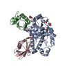
Mass: 94.971 Da / Num. of mol.: 1 / Source method: obtained synthetically / Formula: PO4
#5: Chemical
ChemComp-GOL / GLYCEROL / GLYCERIN / PROPANE-1,2,3-TRIOL


Mass: 92.094 Da / Num. of mol.: 4 / Source method: obtained synthetically / Formula: C3H8O3
#6: Water ChemComp-HOH / water


Mass: 18.015 Da / Num. of mol.: 3 / Source method: isolated from a natural source / Formula: H2O

-
Details

Has protein modificationY

-
Experimental details

-
Experiment

ExperimentMethod: X-RAY DIFFRACTION / Number of used crystals: 1

-
Sample preparation

CrystalDensity Matthews: 2.23 Å3/Da / Density % sol: 44.83 %
Crystal growTemperature: 291 K / Method: vapor diffusion, sitting drop
Details: 0.1 M phosphate citrate (pH 4.2), 20% (w/v) PEG 8K, 0.2 M sodium chloride

-
Data collection

DiffractionMean temperature: 100 K
Diffraction sourceSource: SYNCHROTRON / Site: Diamond / Beamline: I03 / Wavelength: 0.97626 Å
DetectorType: DECTRIS PILATUS3 6M / Detector: PIXEL / Date: Feb 14, 2014
RadiationProtocol: SINGLE WAVELENGTH / Monochromatic (M) / Laue (L): M / Scattering type: x-ray
Radiation wavelengthWavelength: 0.97626 Å / Relative weight: 1
ReflectionResolution: 2.8→56.52 Å / Num. obs: 11505 / % possible obs: 94.6 % / Redundancy: 4.6 % / Rmerge(I) obs: 0.145 / Net I/σ(I): 9.5
Reflection shellResolution: 2.8→2.87 Å / Redundancy: 4.6 % / Rmerge(I) obs: 0.689 / Mean I/σ(I) obs: 2 / % possible all: 95.5

-
Processing

Software
NameVersionClassification
PHENIX1.9_1692refinement
XDSdata reduction
Aimlessdata scaling
PHASERphasing
RefinementMethod to determine structure: MOLECULAR REPLACEMENT / Resolution: 2.8→48.162 Å / SU ML: 0.36 / Cross valid method: FREE R-VALUE / σ(F): 1.36 / Phase error: 23.88
RfactorNum. reflection% reflection
Rfree0.2442 550 4.79 %
Rwork0.2072 --
obs0.209 11478 93.29 %
Solvent computationShrinkage radii: 0.9 Å / VDW probe radii: 1.11 Å
Refinement stepCycle: LAST / Resolution: 2.8→48.162 Å
ProteinNucleic acidLigandSolventTotal
Num. atoms3324 0 29 3 3356
Refine LS restraints
Refine-IDTypeDev idealNumber
X-RAY DIFFRACTIONf_bond_d0.0023422
X-RAY DIFFRACTIONf_angle_d0.5214667
X-RAY DIFFRACTIONf_dihedral_angle_d12.0871215
X-RAY DIFFRACTIONf_chiral_restr0.022538
X-RAY DIFFRACTIONf_plane_restr0.003595
LS refinement shell
Resolution (Å)Rfactor RfreeNum. reflection RfreeRfactor RworkNum. reflection RworkRefine-ID% reflection obs (%)
2.8-3.08170.311420.28172712X-RAY DIFFRACTION95
3.0817-3.52760.28161300.24362738X-RAY DIFFRACTION95
3.5276-4.44390.22711410.18742708X-RAY DIFFRACTION93
4.4439-48.16940.21161370.17622770X-RAY DIFFRACTION91
Refinement TLS params.

Method: refined / Refine-ID: X-RAY DIFFRACTION

IDL112)L122)L132)L222)L232)L332)S11 (Å °)S12 (Å °)S13 (Å °)S21 (Å °)S22 (Å °)S23 (Å °)S31 (Å °)S32 (Å °)S33 (Å °)T112)T122)T132)T222)T232)T332)Origin x (Å)Origin y (Å)Origin z (Å)
12.2536-0.605-0.89621.04070.30751.57330.0478-0.1373-0.1236-0.0119-0.05120.0467-0.05710.0131-0.00060.3174-0.0489-0.03640.21170.0140.238-8.659-0.8852-9.5667
27.2012-4.05010.04085.44071.6792.10370.0161-0.29320.07820.01150.0421-0.2195-0.1765-0.0334-0.01710.405-0.0507-0.00820.2560.02960.202617.4816-17.3895-10.8205
31.0874-0.37251.46514.3056-2.91798.8034-0.3315-1.6781-0.10080.70210.2973-0.1065-0.16980.4570.06980.4950.0908-0.04951.06340.1230.4235-1.8403-5.390117.5648
Refinement TLS group
IDRefine-IDRefine TLS-IDSelection details
1X-RAY DIFFRACTION1chain 'A'
2X-RAY DIFFRACTION2chain 'B'
3X-RAY DIFFRACTION3chain 'C'

+
About Yorodumi

-
News

-
Feb 9, 2022. New format data for meta-information of EMDB entries

New format data for meta-information of EMDB entries

  • Version 3 of the EMDB header file is now the official format.
  • The previous official version 1.9 will be removed from the archive.

Related info.:EMDB header

External links:wwPDB to switch to version 3 of the EMDB data model

-
Aug 12, 2020. Covid-19 info

Covid-19 info

URL: https://pdbj.org/emnavi/covid19.php

New page: Covid-19 featured information page in EM Navigator.

Related info.:Covid-19 info / Mar 5, 2020. Novel coronavirus structure data

+
Mar 5, 2020. Novel coronavirus structure data

Novel coronavirus structure data

Related info.:Yorodumi Speices / Aug 12, 2020. Covid-19 info

External links:COVID-19 featured content - PDBj / Molecule of the Month (242):Coronavirus Proteases

+
Jan 31, 2019. EMDB accession codes are about to change! (news from PDBe EMDB page)

EMDB accession codes are about to change! (news from PDBe EMDB page)

  • The allocation of 4 digits for EMDB accession codes will soon come to an end. Whilst these codes will remain in use, new EMDB accession codes will include an additional digit and will expand incrementally as the available range of codes is exhausted. The current 4-digit format prefixed with “EMD-” (i.e. EMD-XXXX) will advance to a 5-digit format (i.e. EMD-XXXXX), and so on. It is currently estimated that the 4-digit codes will be depleted around Spring 2019, at which point the 5-digit format will come into force.
  • The EM Navigator/Yorodumi systems omit the EMD- prefix.

Related info.:Q: What is EMD? / ID/Accession-code notation in Yorodumi/EM Navigator

External links:EMDB Accession Codes are Changing Soon! / Contact to PDBj

+
Jul 12, 2017. Major update of PDB

Major update of PDB

  • wwPDB released updated PDB data conforming to the new PDBx/mmCIF dictionary.
  • This is a major update changing the version number from 4 to 5, and with Remediation, in which all the entries are updated.
  • In this update, many items about electron microscopy experimental information are reorganized (e.g. em_software).
  • Now, EM Navigator and Yorodumi are based on the updated data.

External links:wwPDB Remediation / Enriched Model Files Conforming to OneDep Data Standards Now Available in the PDB FTP Archive

-
Yorodumi

Thousand views of thousand structures

  • Yorodumi is a browser for structure data from EMDB, PDB, SASBDB, etc.
  • This page is also the successor to EM Navigator detail page, and also detail information page/front-end page for Omokage search.
  • The word "yorodu" (or yorozu) is an old Japanese word meaning "ten thousand". "mi" (miru) is to see.

Related info.:EMDB / PDB / SASBDB / Comparison of 3 databanks / Yorodumi Search / Aug 31, 2016. New EM Navigator & Yorodumi / Yorodumi Papers / Jmol/JSmol / Function and homology information / Changes in new EM Navigator and Yorodumi

Read more