| Entry | Database: PDB / ID: 5lre
|
|---|
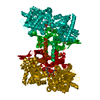
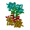
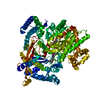
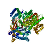
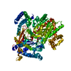
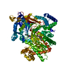
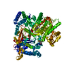
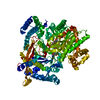
| Title | Crystal structure of Glycogen Phosphorylase b in complex with KS382 |
|---|
 Components Components | Glycogen phosphorylase, muscle form |
|---|
 Keywords Keywords | TRANSFERASE / alpha and beta protein |
|---|
| Function / homology |  Function and homology information Function and homology information
glycogen phosphorylase / glycogen phosphorylase activity / glycogen catabolic process / skeletal muscle myofibril / pyridoxal phosphate binding / nucleotide bindingSimilarity search - Function Glycogen/starch/alpha-glucan phosphorylase / Phosphorylase pyridoxal-phosphate attachment site / Phosphorylase pyridoxal-phosphate attachment site. / Glycosyl transferase, family 35 / Carbohydrate phosphorylase / Glycogen Phosphorylase B; / Rossmann fold / 3-Layer(aba) Sandwich / Alpha BetaSimilarity search - Domain/homology |
|---|
| Biological species |   Oryctolagus cuniculus (rabbit) Oryctolagus cuniculus (rabbit) |
|---|
| Method |  X-RAY DIFFRACTION / SYNCHROTRON / FOURIER SYNTHESIS / Resolution: 1.8 Å X-RAY DIFFRACTION / SYNCHROTRON / FOURIER SYNTHESIS / Resolution: 1.8 Å |
|---|
 Authors Authors | Kantsadi, A.L. / Stravodimos, G.A. / Kyriakis, E. / Chatzileontiadou, D.S.M. / Leonidas, D.D. |
|---|
 Citation Citation | Journal: Eur J Med Chem / Year: 2016
Title: Synthetic, enzyme kinetic, and protein crystallographic studies of C-beta-d-glucopyranosyl pyrroles and imidazoles reveal and explain low nanomolar inhibition of human liver glycogen phosphorylase.
Authors: Kantsadi, A.L. / Bokor, E. / Kun, S. / Stravodimos, G.A. / Chatzileontiadou, D.S. / Leonidas, D.D. / Juhasz-Toth, E. / Szakacs, A. / Batta, G. / Docsa, T. / Gergely, P. / Somsak, L. |
|---|
| History | | Deposition | Aug 18, 2016 | Deposition site: PDBE / Processing site: PDBE |
|---|
| Revision 1.0 | May 31, 2017 | Provider: repository / Type: Initial release |
|---|
| Revision 1.1 | Jan 17, 2018 | Group: Data collection / Category: diffrn_source / Item: _diffrn_source.pdbx_synchrotron_site |
|---|
| Revision 1.2 | Apr 9, 2025 | Group: Advisory / Data collection ...Advisory / Data collection / Database references / Structure summary
Category: chem_comp_atom / chem_comp_bond ...chem_comp_atom / chem_comp_bond / database_2 / pdbx_entry_details / pdbx_unobs_or_zero_occ_atoms
Item: _database_2.pdbx_DOI / _database_2.pdbx_database_accession |
|---|
|
|---|
 Movie
Movie Controller

 Yorodumi
 Open data
 Basic information
 Structure visualization
 Downloads & links
 Other downloads
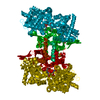


 PDBj
PDBj
 Assembly

 Mass: 357.361 Da / Num. of mol.: 2 / Source method: obtained synthetically / Formula: C18H19N3O5
 Mass: 247.142 Da / Num. of mol.: 1 / Source method: obtained synthetically / Formula: C8H10NO6P
 Mass: 78.133 Da / Num. of mol.: 2 / Source method: obtained synthetically / Formula: C2H6OS / Comment: DMSO, precipitant*YM
 Mass: 18.015 Da / Num. of mol.: 319 / Source method: isolated from a natural source / Formula: H2O
 Sample preparation
 / Beamline: I911-2 / Wavelength: 1.0403 Å
 Processing