 Movie
Movie Controller


+ Open data

- Basic information
| Entry | Database: PDB / ID: 5loo | ||||||||||||
|---|---|---|---|---|---|---|---|---|---|---|---|---|---|
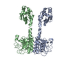
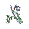
| Title | Structure of full length unliganded CodY from Bacillus subtilis | ||||||||||||
 Components Components | GTP-sensing transcriptional pleiotropic repressor CodY | ||||||||||||
 Keywords Keywords | TRANSCRIPTION / GAF / wHTH / transcriptional regulator / CodY | ||||||||||||
| Function / homology |  Function and homology information Function and homology informationDNA-binding transcription repressor activity / protein-DNA complex / transcription cis-regulatory region binding / regulation of DNA-templated transcription / GTP binding / cytoplasm Similarity search - Function | ||||||||||||
| Biological species |  | ||||||||||||
| Method |  X-RAY DIFFRACTION / SYNCHROTRON / MOLECULAR REPLACEMENT / Resolution: 4.5 Å X-RAY DIFFRACTION / SYNCHROTRON / MOLECULAR REPLACEMENT / Resolution: 4.5 Å | ||||||||||||
 Authors Authors | Wilkinson, A.J. / Levdikov, V.M. / Blagova, E.V. | ||||||||||||
| Funding support |  United Kingdom, United Kingdom,  United States, 3items United States, 3items
| ||||||||||||
 Citation Citation | Journal: J. Biol. Chem. / Year: 2017 Title: Structure of the Branched-chain Amino Acid and GTP-sensing Global Regulator, CodY, from Bacillus subtilis. Authors: Levdikov, V.M. / Blagova, E. / Young, V.L. / Belitsky, B.R. / Lebedev, A. / Sonenshein, A.L. / Wilkinson, A.J. #1: Journal: J. Mol. Biol. / Year: 2009 Title: Structural rearrangement accompanying ligand binding in the GAF domain of CodY from Bacillus subtilis. Authors: Levdikov, V.M. / Blagova, E. / Colledge, V.L. / Lebedev, A.A. / Williamson, D.C. / Sonenshein, A.L. / Wilkinson, A.J. #2: Journal: J. Biol. Chem. / Year: 2006Title: The structure of CodY, a GTP- and isoleucine-responsive regulator of stationary phase and virulence in gram-positive bacteria. Authors: Levdikov, V.M. / Blagova, E. / Joseph, P. / Sonenshein, A.L. / Wilkinson, A.J. | ||||||||||||
| History |
|
- Structure visualization
| Structure viewer | Molecule: MolmilJmol/JSmol |
|---|
- Downloads & links
-Download
| PDBx/mmCIF format | 5loo.cif.gz | 682.3 KB | Display | PDBx/mmCIF format |
|---|---|---|---|---|
| PDB format | pdb5loo.ent.gz | 567.8 KB | Display | PDB format |
| PDBx/mmJSON format | 5loo.json.gz | Tree view | PDBx/mmJSON format | |
| Others |  Other downloads Other downloads |
-Validation report
| Arichive directory | https://data.pdbj.org/pub/pdb/validation_reports/lo/5looftp://data.pdbj.org/pub/pdb/validation_reports/lo/5loo | HTTPS FTP |
|---|
-Related structure data
| Related structure data |  5lnhC  5loeC  5lojSC C: citing same article ( S: Starting model for refinement |
|---|---|
| Similar structure data |


-Links
 PDBj
PDBj

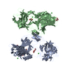
- Assembly
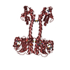
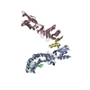
| Deposited unit | 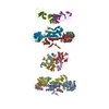
| ||||||||
|---|---|---|---|---|---|---|---|---|---|
| 1 | 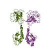
| ||||||||
| 2 | 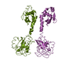
| ||||||||
| 3 | 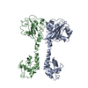
| ||||||||
| 4 | 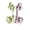
| ||||||||
| 5 | 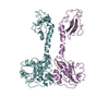
| ||||||||
| 6 | 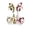
| ||||||||
| 7 | 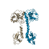
| ||||||||
| 8 | 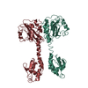
| ||||||||
| Unit cell |
|
-Components
| #1: Protein | Mass: 29604.713 Da / Num. of mol.: 14 Source method: isolated from a genetically manipulated source Source: (gene. exp.) |
|---|
-Experimental details
-Experiment
| Experiment | Method: X-RAY DIFFRACTION / Number of used crystals: 1 |
|---|
- Sample preparation
| Crystal | Density Matthews: 3.4 Å3/Da / Density % sol: 63.1 % |
|---|---|
| Crystal grow | Temperature: 293 K / Method: vapor diffusion, hanging drop / pH: 6 / Details: 1M sodium citrate pH6.0, 5% glycerol |
-Data collection
| Diffraction | Mean temperature: 100 K |
|---|---|
| Diffraction source | Source: SYNCHROTRON / Site: SRS / Beamline: PX14.1 / Wavelength: 0.9804 Å |
| Detector | Type: ADSC QUANTUM 4 / Detector: CCD / Date: Nov 27, 2001 |
| Radiation | Protocol: SINGLE WAVELENGTH / Monochromatic (M) / Laue (L): M / Scattering type: x-ray |
| Radiation wavelength | Wavelength: 0.9804 Å / Relative weight: 1 |
| Reflection | Resolution: 4.5→20 Å / Num. obs: 29671 / % possible obs: 71.4 % / Redundancy: 3.9 % / Rmerge(I) obs: 0.09 / Net I/σ(I): 6.77 |
| Reflection shell | Resolution: 4.5→4.58 Å / Redundancy: 1.3 % / Rmerge(I) obs: 0.508 / Mean I/σ(I) obs: 1.01 / % possible all: 71.4 |
- Processing
| Software |
| ||||||||||||||||||||||||||||||||||||||||||||||||||||||||||||||||||||||||||||||||||||||||||||||||||||||||||||||||||||||||||||||||||||||||||||||||||||||||||||||||||||||||||||||||||||||
|---|---|---|---|---|---|---|---|---|---|---|---|---|---|---|---|---|---|---|---|---|---|---|---|---|---|---|---|---|---|---|---|---|---|---|---|---|---|---|---|---|---|---|---|---|---|---|---|---|---|---|---|---|---|---|---|---|---|---|---|---|---|---|---|---|---|---|---|---|---|---|---|---|---|---|---|---|---|---|---|---|---|---|---|---|---|---|---|---|---|---|---|---|---|---|---|---|---|---|---|---|---|---|---|---|---|---|---|---|---|---|---|---|---|---|---|---|---|---|---|---|---|---|---|---|---|---|---|---|---|---|---|---|---|---|---|---|---|---|---|---|---|---|---|---|---|---|---|---|---|---|---|---|---|---|---|---|---|---|---|---|---|---|---|---|---|---|---|---|---|---|---|---|---|---|---|---|---|---|---|---|---|---|---|
| Refinement | Method to determine structure: MOLECULAR REPLACEMENT Starting model: 5LOJ Resolution: 4.5→19.88 Å / Cor.coef. Fo:Fc: 0.941 / Cor.coef. Fo:Fc free: 0.824 / SU B: 165.968 / SU ML: 2.029 / Cross valid method: THROUGHOUT / ESU R Free: 1.999 / Details: HYDROGENS HAVE BEEN ADDED IN THE RIDING POSITIONS
| ||||||||||||||||||||||||||||||||||||||||||||||||||||||||||||||||||||||||||||||||||||||||||||||||||||||||||||||||||||||||||||||||||||||||||||||||||||||||||||||||||||||||||||||||||||||
| Solvent computation | Ion probe radii: 0.8 Å / Shrinkage radii: 0.8 Å / VDW probe radii: 1.2 Å | ||||||||||||||||||||||||||||||||||||||||||||||||||||||||||||||||||||||||||||||||||||||||||||||||||||||||||||||||||||||||||||||||||||||||||||||||||||||||||||||||||||||||||||||||||||||
| Displacement parameters | Biso mean: 235.091 Å2
| ||||||||||||||||||||||||||||||||||||||||||||||||||||||||||||||||||||||||||||||||||||||||||||||||||||||||||||||||||||||||||||||||||||||||||||||||||||||||||||||||||||||||||||||||||||||
| Refinement step | Cycle: 1 / Resolution: 4.5→19.88 Å
| ||||||||||||||||||||||||||||||||||||||||||||||||||||||||||||||||||||||||||||||||||||||||||||||||||||||||||||||||||||||||||||||||||||||||||||||||||||||||||||||||||||||||||||||||||||||
| Refine LS restraints |
|