 Movie
Movie Controller

[English] 日本語
 Yorodumi
- PDB-6h9o: Complex of the periplasmic domains of bacterial cell division pro... -
+ Open data

- Basic information
| Entry | Database: PDB / ID: 6h9o | |||||||||
|---|---|---|---|---|---|---|---|---|---|---|
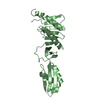
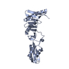
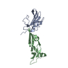
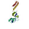
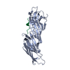
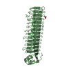
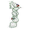
| Title | Complex of the periplasmic domains of bacterial cell division proteins FtsQ and FtsB | |||||||||
 Components Components |
| |||||||||
 Keywords Keywords | CELL CYCLE / bacterial cell division / divisome | |||||||||
| Function / homology |  Function and homology information Function and homology informationFtsQBL complex / divisome complex / division septum assembly / FtsZ-dependent cytokinesis / cell division site / cell division / identical protein binding / plasma membrane Similarity search - Function | |||||||||
| Biological species |  | |||||||||
| Method |  X-RAY DIFFRACTION / SYNCHROTRON / MOLECULAR REPLACEMENT / Resolution: 2.8 Å X-RAY DIFFRACTION / SYNCHROTRON / MOLECULAR REPLACEMENT / Resolution: 2.8 Å | |||||||||
 Authors Authors | Kureisaite-Ciziene, D. / Lowe, J. | |||||||||
| Funding support |  United Kingdom, 2items United Kingdom, 2items
| |||||||||
 Citation Citation | Journal: MBio / Year: 2018 Title: Structural Analysis of the Interaction between the Bacterial Cell Division Proteins FtsQ and FtsB. Authors: Kureisaite-Ciziene, D. / Varadajan, A. / McLaughlin, S.H. / Glas, M. / Monton Silva, A. / Luirink, R. / Mueller, C. / den Blaauwen, T. / Grossmann, T.N. / Luirink, J. / Lowe, J. | |||||||||
| History |
|
- Structure visualization
| Structure viewer | Molecule: MolmilJmol/JSmol |
|---|
- Downloads & links
-Download
| PDBx/mmCIF format | 6h9o.cif.gz | 102.5 KB | Display | PDBx/mmCIF format |
|---|---|---|---|---|
| PDB format | pdb6h9o.ent.gz | 77.8 KB | Display | PDB format |
| PDBx/mmJSON format | 6h9o.json.gz | Tree view | PDBx/mmJSON format | |
| Others |  Other downloads Other downloads |
-Validation report
| Arichive directory | https://data.pdbj.org/pub/pdb/validation_reports/h9/6h9oftp://data.pdbj.org/pub/pdb/validation_reports/h9/6h9o | HTTPS FTP |
|---|
-Related structure data
| Related structure data |  6h9nC  2vh1S S: Starting model for refinement C: citing same article ( |
|---|---|
| Similar structure data |

-Links
 PDBj
PDBj- Assembly
| Deposited unit | 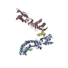
| ||||||||
|---|---|---|---|---|---|---|---|---|---|
| 1 | 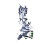
| ||||||||
| 2 | 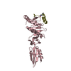
| ||||||||
| Unit cell |
|
-Components
| #1: Protein | Mass: 26269.643 Da / Num. of mol.: 2 Source method: isolated from a genetically manipulated source Source: (gene. exp.) #2: Protein/peptide | Mass: 2883.177 Da / Num. of mol.: 2 / Source method: obtained synthetically Details: Met 77 in FtsB was replaced with Norleucin (Nle) as this is a synthetic peptide Source: (synth.) Has protein modification | Y | |
|---|
-Experimental details
-Experiment
| Experiment | Method: X-RAY DIFFRACTION / Number of used crystals: 1 |
|---|
- Sample preparation
| Crystal | Density Matthews: 2.94 Å3/Da / Density % sol: 58.11 % |
|---|---|
| Crystal grow | Temperature: 293 K / Method: vapor diffusion / pH: 6.5 Details: 0.057 M potassium thiocyanate, 10 % PEG 550 MME, 0.1 M sodium cacodylate pH 6.5. |
-Data collection
| Diffraction | Mean temperature: 100 K |
|---|---|
| Diffraction source | Source: SYNCHROTRON / Site: Diamond / Beamline: I04 / Wavelength: 0.9795 Å |
| Detector | Type: DECTRIS PILATUS3 6M / Detector: PIXEL / Date: Apr 22, 2017 |
| Radiation | Protocol: SINGLE WAVELENGTH / Monochromatic (M) / Laue (L): M / Scattering type: x-ray |
| Radiation wavelength | Wavelength: 0.9795 Å / Relative weight: 1 |
| Reflection | Resolution: 2.8→50 Å / Num. obs: 15922 / % possible obs: 98.5 % / Redundancy: 3.2 % / CC1/2: 0.998 / Rmerge(I) obs: 0.077 / Rpim(I) all: 0.051 / Net I/σ(I): 10.6 |
| Reflection shell | Resolution: 2.8→2.95 Å / Redundancy: 3.3 % / Rmerge(I) obs: 0.744 / Mean I/σ(I) obs: 1.8 / Num. unique obs: 2374 / CC1/2: 0.686 / Rpim(I) all: 0.488 / % possible all: 99.7 |
- Processing
| Software |
| ||||||||||||||||||||||||||||||||||||||||||||||||||||||||||||||||||||||||||||||||||||
|---|---|---|---|---|---|---|---|---|---|---|---|---|---|---|---|---|---|---|---|---|---|---|---|---|---|---|---|---|---|---|---|---|---|---|---|---|---|---|---|---|---|---|---|---|---|---|---|---|---|---|---|---|---|---|---|---|---|---|---|---|---|---|---|---|---|---|---|---|---|---|---|---|---|---|---|---|---|---|---|---|---|---|---|---|---|
| Refinement | Method to determine structure: MOLECULAR REPLACEMENT Starting model: 2VH1 Resolution: 2.8→29.834 Å / SU ML: 0.45 / Cross valid method: FREE R-VALUE / σ(F): 1.14 / Phase error: 30.72
| ||||||||||||||||||||||||||||||||||||||||||||||||||||||||||||||||||||||||||||||||||||
| Solvent computation | Shrinkage radii: 0.9 Å / VDW probe radii: 1.11 Å | ||||||||||||||||||||||||||||||||||||||||||||||||||||||||||||||||||||||||||||||||||||
| Refinement step | Cycle: LAST / Resolution: 2.8→29.834 Å
| ||||||||||||||||||||||||||||||||||||||||||||||||||||||||||||||||||||||||||||||||||||
| Refine LS restraints |
| ||||||||||||||||||||||||||||||||||||||||||||||||||||||||||||||||||||||||||||||||||||
| LS refinement shell |
|
