[English] 日本語
 Yorodumi
- PDB-5lk2: Structure of hantavirus envelope glycoprotein Gc in postfusion co... -
+ Open data
- Basic information
| Entry | Database: PDB / ID: 5lk2 | |||||||||
|---|---|---|---|---|---|---|---|---|---|---|
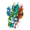
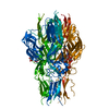
| Title | Structure of hantavirus envelope glycoprotein Gc in postfusion conformation in presence of 300 mM KCL | |||||||||
 Components Components | Envelopment polyprotein | |||||||||
 Keywords Keywords | VIRAL PROTEIN / Hantavirus / Glycoprotein / Viral fusion | |||||||||
| Function / homology |  Function and homology information Function and homology informationsymbiont-mediated suppression of host autophagy / symbiont-mediated suppression of host TRAF-mediated signal transduction / host cell Golgi membrane / host cell mitochondrion / symbiont-mediated suppression of host cytoplasmic pattern recognition receptor signaling pathway via inhibition of MAVS activity / host cell surface / host cell endoplasmic reticulum membrane / endocytosis involved in viral entry into host cell / symbiont-mediated activation of host autophagy / fusion of virus membrane with host endosome membrane ...symbiont-mediated suppression of host autophagy / symbiont-mediated suppression of host TRAF-mediated signal transduction / host cell Golgi membrane / host cell mitochondrion / symbiont-mediated suppression of host cytoplasmic pattern recognition receptor signaling pathway via inhibition of MAVS activity / host cell surface / host cell endoplasmic reticulum membrane / endocytosis involved in viral entry into host cell / symbiont-mediated activation of host autophagy / fusion of virus membrane with host endosome membrane / viral envelope / virion attachment to host cell / virion membrane / signal transduction / zinc ion binding / membrane Similarity search - Function | |||||||||
| Biological species |  Hantaan virus Hantaan virus | |||||||||
| Method |  X-RAY DIFFRACTION / X-RAY DIFFRACTION /  SYNCHROTRON / SYNCHROTRON /  MOLECULAR REPLACEMENT / Resolution: 1.6 Å MOLECULAR REPLACEMENT / Resolution: 1.6 Å | |||||||||
 Authors Authors | Guardado-Calvo, P. / Rey, F.A. | |||||||||
 Citation Citation |  Journal: Plos Pathog. / Year: 2016 Journal: Plos Pathog. / Year: 2016Title: Mechanistic Insight into Bunyavirus-Induced Membrane Fusion from Structure-Function Analyses of the Hantavirus Envelope Glycoprotein Gc. Authors: Guardado-Calvo, P. / Bignon, E.A. / Stettner, E. / Jeffers, S.A. / Perez-Vargas, J. / Pehau-Arnaudet, G. / Tortorici, M.A. / Jestin, J.L. / England, P. / Tischler, N.D. / Rey, F.A. | |||||||||
| History |
|
- Structure visualization
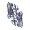
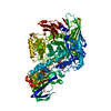
| Structure viewer | Molecule:  Molmil Molmil Jmol/JSmol Jmol/JSmol |
|---|
- Downloads & links
- Download
| PDBx/mmCIF format |  5lk2.cif.gz 5lk2.cif.gz | 175 KB | Display |  PDBx/mmCIF format PDBx/mmCIF format |
|---|---|---|---|---|
| PDB format |  pdb5lk2.ent.gz pdb5lk2.ent.gz | 135.6 KB | Display |  PDB format PDB format |
| PDBx/mmJSON format |  5lk2.json.gz 5lk2.json.gz | Tree view |  PDBx/mmJSON format PDBx/mmJSON format | |
| Others |  Other downloads Other downloads |
-Validation report
| Summary document |  5lk2_validation.pdf.gz 5lk2_validation.pdf.gz | 858.3 KB | Display |  wwPDB validaton report wwPDB validaton report |
|---|---|---|---|---|
| Full document |  5lk2_full_validation.pdf.gz 5lk2_full_validation.pdf.gz | 862.6 KB | Display | |
| Data in XML |  5lk2_validation.xml.gz 5lk2_validation.xml.gz | 19 KB | Display | |
| Data in CIF |  5lk2_validation.cif.gz 5lk2_validation.cif.gz | 28.6 KB | Display | |
| Arichive directory |  https://data.pdbj.org/pub/pdb/validation_reports/lk/5lk2 https://data.pdbj.org/pub/pdb/validation_reports/lk/5lk2 ftp://data.pdbj.org/pub/pdb/validation_reports/lk/5lk2 ftp://data.pdbj.org/pub/pdb/validation_reports/lk/5lk2 | HTTPS FTP |
-Related structure data
| Related structure data |  5ljxC  5ljyC  5ljzSC  5lk0C  5lk1C  5lk3C S: Starting model for refinement C: citing same article ( |
|---|---|
| Similar structure data |
- Links
Links
- Assembly
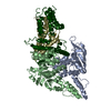
| Deposited unit | 
| ||||||||||||||||||
|---|---|---|---|---|---|---|---|---|---|---|---|---|---|---|---|---|---|---|---|
| 1 | 
| ||||||||||||||||||
| Unit cell |
| ||||||||||||||||||
| Components on special symmetry positions |
|
- Components
| #1: Protein | Mass: 54291.707 Da / Num. of mol.: 1 Source method: isolated from a genetically manipulated source Source: (gene. exp.)  Hantaan virus / Gene: GP / Plasmid: pMT / Cell line (production host): S2 cells / Production host: Hantaan virus / Gene: GP / Plasmid: pMT / Cell line (production host): S2 cells / Production host:  |
|---|---|
| #2: Polysaccharide | 2-acetamido-2-deoxy-beta-D-glucopyranose-(1-4)-[beta-L-fucopyranose-(1-6)]2-acetamido-2-deoxy-beta- ...2-acetamido-2-deoxy-beta-D-glucopyranose-(1-4)-[beta-L-fucopyranose-(1-6)]2-acetamido-2-deoxy-beta-D-glucopyranose Source method: isolated from a genetically manipulated source |
| #3: Chemical | ChemComp-K / |
| #4: Chemical | ChemComp-NA / |
| #5: Water | ChemComp-HOH / |
| Has protein modification | Y |
-Experimental details
-Experiment
| Experiment | Method:  X-RAY DIFFRACTION / Number of used crystals: 1 X-RAY DIFFRACTION / Number of used crystals: 1 |
|---|
- Sample preparation
| Crystal | Density Matthews: 2.62 Å3/Da / Density % sol: 52.97 % |
|---|---|
| Crystal grow | Temperature: 291 K / Method: vapor diffusion, sitting drop / pH: 6.5 Details: 0.1M MES 6.5, 10.77%(v/v) PEG 8000, 7% (v/v) glycerol, 300 mM KCl |
-Data collection
| Diffraction | Mean temperature: 100 K |
|---|---|
| Diffraction source | Source:  SYNCHROTRON / Site: SYNCHROTRON / Site:  ESRF ESRF  / Beamline: ID29 / Wavelength: 0.976251 Å / Beamline: ID29 / Wavelength: 0.976251 Å |
| Detector | Type: DECTRIS PILATUS3 S 6M / Detector: PIXEL / Date: Sep 7, 2013 |
| Radiation | Monochromator: Si(111) / Protocol: SINGLE WAVELENGTH / Monochromatic (M) / Laue (L): M / Scattering type: x-ray |
| Radiation wavelength | Wavelength: 0.976251 Å / Relative weight: 1 |
| Reflection | Resolution: 1.6→37.61 Å / Num. obs: 72553 / % possible obs: 99.7 % / Redundancy: 4.3 % / Biso Wilson estimate: 14.9 Å2 / CC1/2: 0.995 / Rmerge(I) obs: 0.068 / Net I/σ(I): 12.1 |
| Reflection shell | Resolution: 1.6→1.63 Å / Redundancy: 4.3 % / Rmerge(I) obs: 0.343 / Mean I/σ(I) obs: 3.5 / CC1/2: 0.84 / % possible all: 96.8 |
- Processing
| Software |
| ||||||||||||||||||||||||||||||||||||||||||||||||||||||||||||||||||||||||||||||||||||||||||||||||||||||||||||||||||||||||||||||||||||||||||||||||||||||||||||||||||||||||||||||||||||||
|---|---|---|---|---|---|---|---|---|---|---|---|---|---|---|---|---|---|---|---|---|---|---|---|---|---|---|---|---|---|---|---|---|---|---|---|---|---|---|---|---|---|---|---|---|---|---|---|---|---|---|---|---|---|---|---|---|---|---|---|---|---|---|---|---|---|---|---|---|---|---|---|---|---|---|---|---|---|---|---|---|---|---|---|---|---|---|---|---|---|---|---|---|---|---|---|---|---|---|---|---|---|---|---|---|---|---|---|---|---|---|---|---|---|---|---|---|---|---|---|---|---|---|---|---|---|---|---|---|---|---|---|---|---|---|---|---|---|---|---|---|---|---|---|---|---|---|---|---|---|---|---|---|---|---|---|---|---|---|---|---|---|---|---|---|---|---|---|---|---|---|---|---|---|---|---|---|---|---|---|---|---|---|---|
| Refinement | Method to determine structure:  MOLECULAR REPLACEMENT MOLECULAR REPLACEMENTStarting model: 5LJZ Resolution: 1.6→37.61 Å / SU ML: 0.17 / Cross valid method: FREE R-VALUE / σ(F): 1.97 / Phase error: 18.99
| ||||||||||||||||||||||||||||||||||||||||||||||||||||||||||||||||||||||||||||||||||||||||||||||||||||||||||||||||||||||||||||||||||||||||||||||||||||||||||||||||||||||||||||||||||||||
| Solvent computation | Shrinkage radii: 0.9 Å / VDW probe radii: 1.11 Å | ||||||||||||||||||||||||||||||||||||||||||||||||||||||||||||||||||||||||||||||||||||||||||||||||||||||||||||||||||||||||||||||||||||||||||||||||||||||||||||||||||||||||||||||||||||||
| Refinement step | Cycle: LAST / Resolution: 1.6→37.61 Å
| ||||||||||||||||||||||||||||||||||||||||||||||||||||||||||||||||||||||||||||||||||||||||||||||||||||||||||||||||||||||||||||||||||||||||||||||||||||||||||||||||||||||||||||||||||||||
| Refine LS restraints |
| ||||||||||||||||||||||||||||||||||||||||||||||||||||||||||||||||||||||||||||||||||||||||||||||||||||||||||||||||||||||||||||||||||||||||||||||||||||||||||||||||||||||||||||||||||||||
| LS refinement shell |
|
 Movie
Movie Controller



 PDBj
PDBj


