[English] 日本語
Yorodumi
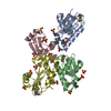
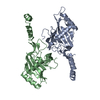
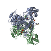
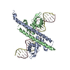
- PDB-5l80: Structure of Exuperantia EXO-like and SAM-like domains -

+
Open data


ID or keywords:

Loading...

-
Basic information

Entry
Database: PDB / ID: 5l80
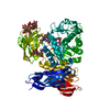
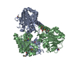
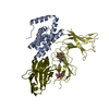
TitleStructure of Exuperantia EXO-like and SAM-like domains
ComponentsMaternal protein exuperantia,Maternal protein exuperantia
KeywordsRNA BINDING PROTEIN / Exonuclease / RNA / pseudo nuclease / SAM domain
Function / homology
Function and homology information


ovarian nurse cell to oocyte transport / embryonic development via the syncytial blastoderm / bicoid mRNA localization / regulation of pole plasm oskar mRNA localization / pole plasm oskar mRNA localization / anterior/posterior axis specification, embryo / precatalytic spliceosome / P-body / mRNA splicing, via spliceosome / cytoplasmic ribonucleoprotein granule ...ovarian nurse cell to oocyte transport / embryonic development via the syncytial blastoderm / bicoid mRNA localization / regulation of pole plasm oskar mRNA localization / pole plasm oskar mRNA localization / anterior/posterior axis specification, embryo / precatalytic spliceosome / P-body / mRNA splicing, via spliceosome / cytoplasmic ribonucleoprotein granule / spermatogenesis / single-stranded RNA binding / protein homodimerization activity / cytoplasm
Similarity search - Function
Maternal protein exuperantia / Exuperantia, SAM-like domain / Exuperantia SAM-like domain / Ribonuclease H superfamily
Similarity search - Domain/homology
Maternal protein exuperantia
Similarity search - Component
Biological speciesDrosophila melanogaster (fruit fly)
MethodX-RAY DIFFRACTION / SYNCHROTRON / MOLECULAR REPLACEMENT / Resolution: 2.8 Å
AuthorsLazzaretti, D. / Veith, K. / Bono, F.
CitationJournal: Nat.Struct.Mol.Biol. / Year: 2016
Title: The bicoid mRNA localization factor Exuperantia is an RNA-binding pseudonuclease.
Authors: Lazzaretti, D. / Veith, K. / Kramer, K. / Basquin, C. / Urlaub, H. / Irion, U. / Bono, F.
History
DepositionJun 5, 2016Deposition site: PDBE / Processing site: PDBE
Revision 1.0Jul 6, 2016Provider: repository / Type: Initial release
Revision 1.1Jul 13, 2016Group: Database references
Revision 1.2Aug 17, 2016Group: Database references
Revision 1.3Jan 10, 2024Group: Data collection / Database references / Refinement description
Category: chem_comp_atom / chem_comp_bond ...chem_comp_atom / chem_comp_bond / database_2 / pdbx_initial_refinement_model
Item: _database_2.pdbx_DOI / _database_2.pdbx_database_accession

-
Structure visualization

Structure viewerMolecule:
MolmilJmol/JSmol

Downloads & links

-
Assembly

Deposited unit
A: Maternal protein exuperantia,Maternal protein exuperantia
B: Maternal protein exuperantia,Maternal protein exuperantia


Theoretical massNumber of molelcules
Total (without water)79,4012
Polymers79,4012
Non-polymers00
Water19811
1


  • Idetical with deposited unit
  • defined by author&software
TypeNameSymmetry operationNumber
identity operation1_555x,y,z1
Buried area2770 Å2
ΔGint-23 kcal/mol
Surface area27410 Å2
MethodPISA
Unit cell
Length a, b, c (Å)79.540, 66.590, 81.330
Angle α, β, γ (deg.)90.00, 113.64, 90.00
Int Tables number4
Space group name H-MP1211

-
Components

#1: Protein Maternal protein exuperantia,Maternal protein exuperantia


Mass: 39700.672 Da / Num. of mol.: 2
Source method: isolated from a genetically manipulated source
Source: (gene. exp.) Drosophila melanogaster (fruit fly) / Gene: exu, CG8994 / Production host: Escherichia coli (E. coli) / References: UniProt: P28750
#2: Water ChemComp-HOH / water


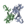
Mass: 18.015 Da / Num. of mol.: 11 / Source method: isolated from a natural source / Formula: H2O

-
Experimental details

-
Experiment

ExperimentMethod: X-RAY DIFFRACTION / Number of used crystals: 1

-
Sample preparation

CrystalDensity Matthews: 2.48 Å3/Da / Density % sol: 50.5 %
Crystal growTemperature: 277 K / Method: vapor diffusion, sitting drop / pH: 6 / Details: Sodium Malonate, PEG 1000, MES

-
Data collection

DiffractionMean temperature: 100 K
Diffraction sourceSource: SYNCHROTRON / Site: SLS / Beamline: X06SA / Wavelength: 1 Å
DetectorType: DECTRIS PILATUS 6M / Detector: PIXEL / Date: Oct 27, 2013
RadiationProtocol: SINGLE WAVELENGTH / Monochromatic (M) / Laue (L): M / Scattering type: x-ray
Radiation wavelengthWavelength: 1 Å / Relative weight: 1
ReflectionResolution: 2.8→50 Å / Num. obs: 19360 / % possible obs: 99.5 % / Redundancy: 6.7 % / Rmerge(I) obs: 0.078 / Net I/σ(I): 19.4
Reflection shellResolution: 2.8→2.9 Å / Redundancy: 6.6 % / Rmerge(I) obs: 1.045 / Mean I/σ(I) obs: 2.2 / % possible all: 99.2

-
Processing

Software
NameVersionClassification
PHENIX1.9_1692refinement
XDSdata reduction
XSCALEdata scaling
PHASERphasing
RefinementMethod to determine structure: MOLECULAR REPLACEMENT
Starting model: 5L7Z
Resolution: 2.8→47.336 Å / SU ML: 0.43 / Cross valid method: FREE R-VALUE / σ(F): 1.36 / Phase error: 32.02
RfactorNum. reflection% reflection
Rfree0.2765 969 5.01 %
Rwork0.2523 --
obs0.2535 19360 99.69 %
Solvent computationShrinkage radii: 0.9 Å / VDW probe radii: 1.11 Å
Refinement stepCycle: LAST / Resolution: 2.8→47.336 Å
ProteinNucleic acidLigandSolventTotal
Num. atoms4229 0 0 11 4240
Refine LS restraints
Refine-IDTypeDev idealNumber
X-RAY DIFFRACTIONf_bond_d0.0034294
X-RAY DIFFRACTIONf_angle_d0.6585823
X-RAY DIFFRACTIONf_dihedral_angle_d11.7411485
X-RAY DIFFRACTIONf_chiral_restr0.027693
X-RAY DIFFRACTIONf_plane_restr0.002743
LS refinement shell
Resolution (Å)Rfactor RfreeNum. reflection RfreeRfactor RworkNum. reflection RworkRefine-ID% reflection obs (%)
2.8-2.94760.36761370.31452600X-RAY DIFFRACTION99
2.9476-3.13230.35431380.30652616X-RAY DIFFRACTION100
3.1323-3.37410.29471360.29742592X-RAY DIFFRACTION100
3.3741-3.71350.28441380.26312622X-RAY DIFFRACTION100
3.7135-4.25050.2741380.23352611X-RAY DIFFRACTION100
4.2505-5.3540.24531390.22432652X-RAY DIFFRACTION100
5.354-47.30.26551430.24792698X-RAY DIFFRACTION100
Refinement TLS params.

Method: refined / Refine-ID: X-RAY DIFFRACTION

IDL112)L122)L132)L222)L232)L332)S11 (Å °)S12 (Å °)S13 (Å °)S21 (Å °)S22 (Å °)S23 (Å °)S31 (Å °)S32 (Å °)S33 (Å °)T112)T122)T132)T222)T232)T332)Origin x (Å)Origin y (Å)Origin z (Å)
13.7696-0.5839-1.57276.56311.17515.4759-0.12-0.009-0.44270.35660.02340.54510.4809-0.23310.04340.45190.00860.03430.35440.04760.4713-25.5027-21.197-11.062
20.97220.1921-0.60291.392-2.29755.52550.4695-0.6938-0.79390.11990.0663-0.26470.4638-0.7855-0.18210.9197-0.1124-0.16050.90190.20520.9874-34.86185.7499-33.2847
31.4502-2.2631-0.30415.14222.52912.7174-0.0795-0.93890.09360.9976-0.08481.0225-1.2403-0.8514-0.3361.31430.1818-0.01220.97660.15240.9085-41.4918.7445-30.7237
44.68241.1299-0.34254.9491-1.01993.8267-0.0079-0.12820.00160.038-0.2233-0.7303-0.41851.06090.11910.6417-0.1462-0.01320.65950.05720.6491-3.50588.9962-18.8851
54.004-0.1891.40368.3511-1.01650.59961.04890.57041.0419-0.8727-0.457-0.711-0.887-1.80920.23441.58560.77750.04771.78810.00610.9395-15.35574.420115.7894
Refinement TLS group
IDRefine-IDRefine TLS-IDSelection details
1X-RAY DIFFRACTION1chain 'A' and (resid 26 through 268 )
2X-RAY DIFFRACTION2chain 'A' and (resid 269 through 329 )
3X-RAY DIFFRACTION3chain 'A' and (resid 330 through 344 )
4X-RAY DIFFRACTION4chain 'B' and (resid 28 through 261 )
5X-RAY DIFFRACTION5chain 'B' and (resid 262 through 343 )

+
About Yorodumi

-
News

-
Feb 9, 2022. New format data for meta-information of EMDB entries

New format data for meta-information of EMDB entries

  • Version 3 of the EMDB header file is now the official format.
  • The previous official version 1.9 will be removed from the archive.

Related info.:EMDB header

External links:wwPDB to switch to version 3 of the EMDB data model

-
Aug 12, 2020. Covid-19 info

Covid-19 info

URL: https://pdbjlvh1.pdbj.org/emnavi/covid19.php

New page: Covid-19 featured information page in EM Navigator.

Related info.:Covid-19 info / Mar 5, 2020. Novel coronavirus structure data

+
Mar 5, 2020. Novel coronavirus structure data

Novel coronavirus structure data

Related info.:Yorodumi Speices / Aug 12, 2020. Covid-19 info

External links:COVID-19 featured content - PDBj / Molecule of the Month (242):Coronavirus Proteases

+
Jan 31, 2019. EMDB accession codes are about to change! (news from PDBe EMDB page)

EMDB accession codes are about to change! (news from PDBe EMDB page)

  • The allocation of 4 digits for EMDB accession codes will soon come to an end. Whilst these codes will remain in use, new EMDB accession codes will include an additional digit and will expand incrementally as the available range of codes is exhausted. The current 4-digit format prefixed with “EMD-” (i.e. EMD-XXXX) will advance to a 5-digit format (i.e. EMD-XXXXX), and so on. It is currently estimated that the 4-digit codes will be depleted around Spring 2019, at which point the 5-digit format will come into force.
  • The EM Navigator/Yorodumi systems omit the EMD- prefix.

Related info.:Q: What is EMD? / ID/Accession-code notation in Yorodumi/EM Navigator

External links:EMDB Accession Codes are Changing Soon! / Contact to PDBj

+
Jul 12, 2017. Major update of PDB

Major update of PDB

  • wwPDB released updated PDB data conforming to the new PDBx/mmCIF dictionary.
  • This is a major update changing the version number from 4 to 5, and with Remediation, in which all the entries are updated.
  • In this update, many items about electron microscopy experimental information are reorganized (e.g. em_software).
  • Now, EM Navigator and Yorodumi are based on the updated data.

External links:wwPDB Remediation / Enriched Model Files Conforming to OneDep Data Standards Now Available in the PDB FTP Archive

-
Yorodumi

Thousand views of thousand structures

  • Yorodumi is a browser for structure data from EMDB, PDB, SASBDB, etc.
  • This page is also the successor to EM Navigator detail page, and also detail information page/front-end page for Omokage search.
  • The word "yorodu" (or yorozu) is an old Japanese word meaning "ten thousand". "mi" (miru) is to see.

Related info.:EMDB / PDB / SASBDB / Comparison of 3 databanks / Yorodumi Search / Aug 31, 2016. New EM Navigator & Yorodumi / Yorodumi Papers / Jmol/JSmol / Function and homology information / Changes in new EM Navigator and Yorodumi

Read more