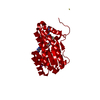
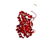
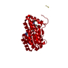
17-beta-hydroxysteroiddehydrogenase14 / 17-beta-HSD 14 / 17-beta-hydroxysteroid dehydrogenase DHRS10 / Dehydrogenase/reductase SDR family ...17-beta-HSD 14 / 17-beta-hydroxysteroid dehydrogenase DHRS10 / Dehydrogenase/reductase SDR family member 10 / Retinal short-chain dehydrogenase/reductase retSDR3 / Short chain dehydrogenase/reductase family 47C member 1
Mass: 28682.740 Da / Num. of mol.: 1 Source method: isolated from a genetically manipulated source Source: (gene. exp.) Homo sapiens (human) / Gene: HSD17B14, DHRS10, SDR3, SDR47C1, UNQ502/PRO474 / Production host: Escherichia coli BL21(DE3) (bacteria) / Variant (production host): pLysS References: UniProt: Q9BPX1, Oxidoreductases; Acting on the CH-OH group of donors; With NAD+ or NADP+ as acceptor
In the structure databanks used in Yorodumi, some data are registered as the other names, "COVID-19 virus" and "2019-nCoV". Here are the details of the virus and the list of structure data.
Jan 31, 2019. EMDB accession codes are about to change! (news from PDBe EMDB page)
EMDB accession codes are about to change! (news from PDBe EMDB page)
The allocation of 4 digits for EMDB accession codes will soon come to an end. Whilst these codes will remain in use, new EMDB accession codes will include an additional digit and will expand incrementally as the available range of codes is exhausted. The current 4-digit format prefixed with “EMD-” (i.e. EMD-XXXX) will advance to a 5-digit format (i.e. EMD-XXXXX), and so on. It is currently estimated that the 4-digit codes will be depleted around Spring 2019, at which point the 5-digit format will come into force.
The EM Navigator/Yorodumi systems omit the EMD- prefix.
Related info.:Q: What is EMD? / ID/Accession-code notation in Yorodumi/EM Navigator
Yorodumi is a browser for structure data from EMDB, PDB, SASBDB, etc.
This page is also the successor to EM Navigator detail page, and also detail information page/front-end page for Omokage search.
The word "yorodu" (or yorozu) is an old Japanese word meaning "ten thousand". "mi" (miru) is to see.
Related info.:EMDB / PDB / SASBDB / Comparison of 3 databanks / Yorodumi Search / Aug 31, 2016. New EM Navigator & Yorodumi / Yorodumi Papers / Jmol/JSmol / Function and homology information / Changes in new EM Navigator and Yorodumi
 Movie
Movie Controller

 Yorodumi
 Open data
 Basic information
 Components
 Keywords
 Function and homology information
 Homo sapiens (human)
 X-RAY DIFFRACTION /
  Authors
 Germany, 2items
  Citation
 Structure visualization
 Downloads & links
 Other downloads
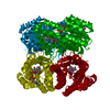


 PDBj
PDBj


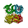
 Assembly


 Type: D-saccharide, beta linking / Mass: 180.156 Da / Num. of mol.: 1


 Mass: 663.425 Da / Num. of mol.: 1 / Source method: obtained synthetically / Formula: C21H27N7O14P2 / Comment: NAD*YM
 Mass: 22.990 Da / Num. of mol.: 1 / Source method: obtained synthetically / Formula: Na
 Mass: 343.281 Da / Num. of mol.: 1 / Source method: obtained synthetically / Formula: C18H11F2NO4
 Sample preparation
 / Beamline: 5.2R / Wavelength: 1 Å
 Processing