 Movie
Movie Controller

[English] 日本語
 Yorodumi
- PDB-5l59: Plexin A1 full extracellular region, domains 1 to 10, to 6 angstr... -
+ Open data

- Basic information
| Entry | Database: PDB / ID: 5l59 | |||||||||
|---|---|---|---|---|---|---|---|---|---|---|
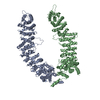
| Title | Plexin A1 full extracellular region, domains 1 to 10, to 6 angstrom, spacegroup P2(1) | |||||||||
 Components Components | Plexin-A1 | |||||||||
 Keywords Keywords | SIGNALING PROTEIN / receptor / signaling / axon guidance | |||||||||
| Function / homology |  Function and homology information Function and homology informationOther semaphorin interactions / olfactory nerve formation / : / CRMPs in Sema3A signaling / neuron projection guidance / Sema3A PAK dependent Axon repulsion / dichotomous subdivision of terminal units involved in salivary gland branching / RND1 GTPase cycle / gonadotrophin-releasing hormone neuronal migration to the hypothalamus / T cell activation via T cell receptor contact with antigen bound to MHC molecule on antigen presenting cell ...Other semaphorin interactions / olfactory nerve formation / : / CRMPs in Sema3A signaling / neuron projection guidance / Sema3A PAK dependent Axon repulsion / dichotomous subdivision of terminal units involved in salivary gland branching / RND1 GTPase cycle / gonadotrophin-releasing hormone neuronal migration to the hypothalamus / T cell activation via T cell receptor contact with antigen bound to MHC molecule on antigen presenting cell / SEMA3A-Plexin repulsion signaling by inhibiting Integrin adhesion / semaphorin receptor complex / semaphorin receptor activity / regulation of smooth muscle cell migration / neuron projection extension / semaphorin-plexin signaling pathway / synapse assembly / regulation of cell migration / synapse / glutamatergic synapse / nucleoplasm / membrane / plasma membrane / cytosol Similarity search - Function | |||||||||
| Biological species |  | |||||||||
| Method |  X-RAY DIFFRACTION / SYNCHROTRON / MOLECULAR REPLACEMENT / Resolution: 6 Å X-RAY DIFFRACTION / SYNCHROTRON / MOLECULAR REPLACEMENT / Resolution: 6 Å | |||||||||
 Authors Authors | Janssen, B.J.C. / Kong, Y. / Malinauskas, T. / Vangoor, V.R. / Coles, C.H. / Kaufmann, R. / Ni, T. / Gilbert, R.J.C. / Padilla-Parra, S. / Pasterkamp, R.J. / Jones, E.Y. | |||||||||
 Citation Citation | Journal: Neuron / Year: 2016 Title: Structural Basis for Plexin Activation and Regulation. Authors: Kong, Y. / Janssen, B.J. / Malinauskas, T. / Vangoor, V.R. / Coles, C.H. / Kaufmann, R. / Ni, T. / Gilbert, R.J. / Padilla-Parra, S. / Pasterkamp, R.J. / Jones, E.Y. | |||||||||
| History |
|
- Structure visualization
| Structure viewer | Molecule: MolmilJmol/JSmol |
|---|
- Downloads & links
-Download
| PDBx/mmCIF format | 5l59.cif.gz | 437.3 KB | Display | PDBx/mmCIF format |
|---|---|---|---|---|
| PDB format | pdb5l59.ent.gz | 339.1 KB | Display | PDB format |
| PDBx/mmJSON format | 5l59.json.gz | Tree view | PDBx/mmJSON format | |
| Others |  Other downloads Other downloads |
-Validation report
| Arichive directory | https://data.pdbj.org/pub/pdb/validation_reports/l5/5l59ftp://data.pdbj.org/pub/pdb/validation_reports/l5/5l59 | HTTPS FTP |
|---|
-Related structure data
| Related structure data |  5l56SC  5l5cC 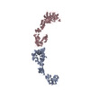 5l5gC  5l5kC  5l5lC  5l5mC 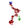 5l5nC 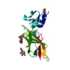 5l74C 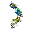 5l7nC S: Starting model for refinement C: citing same article ( |
|---|---|
| Similar structure data |


-Links
 PDBj
PDBj


- Assembly
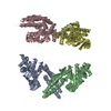
| Deposited unit | 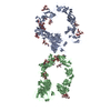
| ||||||||
|---|---|---|---|---|---|---|---|---|---|
| 1 | 
| ||||||||
| 2 | 
| ||||||||
| Unit cell |
|
-Components
-Protein , 1 types, 2 molecules AB
| #1: Protein | Mass: 134024.828 Da / Num. of mol.: 2 / Fragment: UNP residues 37-1236 Source method: isolated from a genetically manipulated source Source: (gene. exp.)  Homo sapiens (human) / References: UniProt: P70206 Homo sapiens (human) / References: UniProt: P70206 |
|---|
-Sugars , 6 types, 20 molecules 
| #2: Polysaccharide | alpha-D-mannopyranose-(1-3)-beta-D-mannopyranose-(1-4)-2-acetamido-2-deoxy-beta-D-glucopyranose-(1- ...alpha-D-mannopyranose-(1-3)-beta-D-mannopyranose-(1-4)-2-acetamido-2-deoxy-beta-D-glucopyranose-(1-4)-2-acetamido-2-deoxy-beta-D-glucopyranose Source method: isolated from a genetically manipulated source #3: Polysaccharide | Source method: isolated from a genetically manipulated source #4: Polysaccharide | alpha-D-mannopyranose-(1-3)-[alpha-D-mannopyranose-(1-6)]alpha-D-mannopyranose-(1-6)-[alpha-D- ...alpha-D-mannopyranose-(1-3)-[alpha-D-mannopyranose-(1-6)]alpha-D-mannopyranose-(1-6)-[alpha-D-mannopyranose-(1-3)]beta-D-mannopyranose-(1-4)-2-acetamido-2-deoxy-beta-D-glucopyranose-(1-4)-2-acetamido-2-deoxy-beta-D-glucopyranose Source method: isolated from a genetically manipulated source #5: Polysaccharide | Source method: isolated from a genetically manipulated source #6: Polysaccharide | Source method: isolated from a genetically manipulated source #7: Sugar | ChemComp-NAG /  Type: D-saccharide, beta linking / Mass: 221.208 Da / Num. of mol.: 4 Type: D-saccharide, beta linking / Mass: 221.208 Da / Num. of mol.: 4Source method: isolated from a genetically manipulated source Formula: C8H15NO6 |
|---|
-Details
| Has protein modification | Y |
|---|
-Experimental details
-Experiment
| Experiment | Method: X-RAY DIFFRACTION / Number of used crystals: 1 |
|---|
- Sample preparation
| Crystal | Density % sol: 80 % |
|---|---|
| Crystal grow | Temperature: 293.5 K / Method: vapor diffusion, sitting drop / pH: 6 Details: 15% (v/v) propanol, 20 mM magnesium chloride 50 mM MES, pH 6.0 |
-Data collection
| Diffraction | Mean temperature: 100 K |
|---|---|
| Diffraction source | Source: SYNCHROTRON / Site: Diamond  / Beamline: I03 / Wavelength: 1.0719 Å / Beamline: I03 / Wavelength: 1.0719 Å |
| Detector | Type: DECTRIS PILATUS 6M-F / Detector: PIXEL / Date: Apr 20, 2011 |
| Radiation | Protocol: SINGLE WAVELENGTH / Monochromatic (M) / Laue (L): M / Scattering type: x-ray |
| Radiation wavelength | Wavelength: 1.0719 Å / Relative weight: 1 |
| Reflection | Resolution: 6→58.218 Å / Num. obs: 19391 / % possible obs: 99.7 % / Redundancy: 3.2 % / Net I/σ(I): 6.8 |
- Processing
| Software |
| ||||||||||||||||||||||||||||||||||||||||||||||||||||||||||||||||||||||||||||||||||||||||||||||||||
|---|---|---|---|---|---|---|---|---|---|---|---|---|---|---|---|---|---|---|---|---|---|---|---|---|---|---|---|---|---|---|---|---|---|---|---|---|---|---|---|---|---|---|---|---|---|---|---|---|---|---|---|---|---|---|---|---|---|---|---|---|---|---|---|---|---|---|---|---|---|---|---|---|---|---|---|---|---|---|---|---|---|---|---|---|---|---|---|---|---|---|---|---|---|---|---|---|---|---|---|
| Refinement | Method to determine structure: MOLECULAR REPLACEMENT Starting model: 5L56 Resolution: 6→58.218 Å / SU ML: 0.79 / Cross valid method: FREE R-VALUE / σ(F): 0.61 / Phase error: 33.24
| ||||||||||||||||||||||||||||||||||||||||||||||||||||||||||||||||||||||||||||||||||||||||||||||||||
| Solvent computation | Shrinkage radii: 0.9 Å / VDW probe radii: 1.11 Å | ||||||||||||||||||||||||||||||||||||||||||||||||||||||||||||||||||||||||||||||||||||||||||||||||||
| Refinement step | Cycle: LAST / Resolution: 6→58.218 Å
| ||||||||||||||||||||||||||||||||||||||||||||||||||||||||||||||||||||||||||||||||||||||||||||||||||
| Refine LS restraints |
| ||||||||||||||||||||||||||||||||||||||||||||||||||||||||||||||||||||||||||||||||||||||||||||||||||
| LS refinement shell |
|
