 Movie
Movie Controller

[English] 日本語
 Yorodumi
- PDB-5kzd: N-acetylneuraminate lyase from methicillin-resistant Staphylococc... -
+ Open data

- Basic information
| Entry | Database: PDB / ID: 5kzd | ||||||
|---|---|---|---|---|---|---|---|
| Title | N-acetylneuraminate lyase from methicillin-resistant Staphylococcus aureus with bound sialic acid alditol | ||||||
 Components Components | N-acetylneuraminate lyase | ||||||
 Keywords Keywords | LYASE / TIM-barrel / inhibitor / N-acetylneuraminate lyase | ||||||
| Function / homology |  Function and homology information Function and homology informationN-acetylneuraminate lyase / N-acetylneuraminate lyase activity / N-acetylneuraminate catabolic process / carbohydrate metabolic process / cytosol Similarity search - Function | ||||||
| Biological species |   Staphylococcus aureus (bacteria) Staphylococcus aureus (bacteria) | ||||||
| Method |  X-RAY DIFFRACTION / SYNCHROTRON / Resolution: 2.334 Å X-RAY DIFFRACTION / SYNCHROTRON / Resolution: 2.334 Å | ||||||
 Authors Authors | North, R.A. / Watson, A.J.A. / Pearce, F.G. / Muscroft-Taylor, A.C. / Friemann, R. / Fairbanks, A.J. / Dobson, R.C.J. | ||||||
 Citation Citation | Journal: FEBS Lett. / Year: 2016 Title: Structure and inhibition of N-acetylneuraminate lyase from methicillin-resistant Staphylococcus aureus. Authors: North, R.A. / Watson, A.J. / Pearce, F.G. / Muscroft-Taylor, A.C. / Friemann, R. / Fairbanks, A.J. / Dobson, R.C. | ||||||
| History |
|
- Structure visualization
| Structure viewer | Molecule: MolmilJmol/JSmol |
|---|
- Downloads & links
-Download
| PDBx/mmCIF format | 5kzd.cif.gz | 231.7 KB | Display | PDBx/mmCIF format |
|---|---|---|---|---|
| PDB format | pdb5kzd.ent.gz | 188.6 KB | Display | PDB format |
| PDBx/mmJSON format | 5kzd.json.gz | Tree view | PDBx/mmJSON format | |
| Others |  Other downloads Other downloads |
-Validation report
| Summary document | 5kzd_validation.pdf.gz | 1.5 MB | Display | wwPDB validaton report |
|---|---|---|---|---|
| Full document | 5kzd_full_validation.pdf.gz | 1.5 MB | Display | |
| Data in XML | 5kzd_validation.xml.gz | 42.7 KB | Display | |
| Data in CIF | 5kzd_validation.cif.gz | 58.7 KB | Display | |
| Arichive directory | https://data.pdbj.org/pub/pdb/validation_reports/kz/5kzdftp://data.pdbj.org/pub/pdb/validation_reports/kz/5kzd | HTTPS FTP |
-Related structure data


-Links
 PDBj
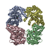
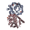
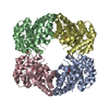
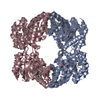
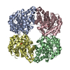
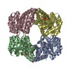
PDBj- Assembly
| Deposited unit | 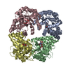
| ||||||||
|---|---|---|---|---|---|---|---|---|---|
| 1 |
| ||||||||
| Unit cell |
|
-Components
| #1: Protein | Mass: 33076.465 Da / Num. of mol.: 4 Source method: isolated from a genetically manipulated source Source: (gene. exp.) Staphylococcus aureus (strain USA300) (bacteria)Strain: USA300 / Gene: nanA, SAUSA300_0315 / Production host: References: UniProt: Q2FJU9, UniProt: Q2G160*PLUS, N-acetylneuraminate lyase #2: Chemical | ChemComp-RCJ / ( 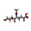  Mass: 311.286 Da / Num. of mol.: 4 / Source method: obtained synthetically / Formula: C11H21NO9 Mass: 311.286 Da / Num. of mol.: 4 / Source method: obtained synthetically / Formula: C11H21NO9#3: Water | ChemComp-HOH / |  Mass: 18.015 Da / Num. of mol.: 177 / Source method: isolated from a natural source / Formula: H2O Mass: 18.015 Da / Num. of mol.: 177 / Source method: isolated from a natural source / Formula: H2O |
|---|
-Experimental details
-Experiment
| Experiment | Method: X-RAY DIFFRACTION / Number of used crystals: 1 |
|---|
- Sample preparation
| Crystal | Density Matthews: 2.2 Å3/Da / Density % sol: 44.17 % |
|---|---|
| Crystal grow | Temperature: 293 K / Method: vapor diffusion, hanging drop / pH: 5.5 Details: PEG 3350, sodium chloride, Tris, sialic acid alditols |
-Data collection
| Diffraction | Mean temperature: 110 K | ||||||||||||||||||
|---|---|---|---|---|---|---|---|---|---|---|---|---|---|---|---|---|---|---|---|
| Diffraction source | Source: SYNCHROTRON / Site: Australian Synchrotron  / Beamline: MX2 / Wavelength: 0.9537 Å / Beamline: MX2 / Wavelength: 0.9537 Å | ||||||||||||||||||
| Detector | Type: ADSC QUANTUM 315r / Detector: CCD / Date: Apr 9, 2015 | ||||||||||||||||||
| Radiation | Protocol: SINGLE WAVELENGTH / Monochromatic (M) / Laue (L): M / Scattering type: x-ray | ||||||||||||||||||
| Radiation wavelength | Wavelength: 0.9537 Å / Relative weight: 1 | ||||||||||||||||||
| Reflection | Resolution: 2.33→38.179 Å / Num. obs: 49980 / % possible obs: 99.1 % / Redundancy: 7.3 % / Biso Wilson estimate: 35.67 Å2 / CC1/2: 0.998 / Rmerge(I) obs: 0.115 / Net I/σ(I): 14.2 | ||||||||||||||||||
| Reflection shell |
|
- Processing
| Software |
| |||||||||||||||||||||||||||||||||||||||||||||||||||||||||||||||||||||||||||||||||||||||||||||||||||||||||
|---|---|---|---|---|---|---|---|---|---|---|---|---|---|---|---|---|---|---|---|---|---|---|---|---|---|---|---|---|---|---|---|---|---|---|---|---|---|---|---|---|---|---|---|---|---|---|---|---|---|---|---|---|---|---|---|---|---|---|---|---|---|---|---|---|---|---|---|---|---|---|---|---|---|---|---|---|---|---|---|---|---|---|---|---|---|---|---|---|---|---|---|---|---|---|---|---|---|---|---|---|---|---|---|---|---|---|
| Refinement | Resolution: 2.334→38.179 Å / SU ML: 0.36 / Cross valid method: FREE R-VALUE / σ(F): 1.34 / Phase error: 29.38
| |||||||||||||||||||||||||||||||||||||||||||||||||||||||||||||||||||||||||||||||||||||||||||||||||||||||||
| Solvent computation | Shrinkage radii: 0.9 Å / VDW probe radii: 1.11 Å | |||||||||||||||||||||||||||||||||||||||||||||||||||||||||||||||||||||||||||||||||||||||||||||||||||||||||
| Displacement parameters | Biso max: 91.1 Å2 / Biso mean: 36.4025 Å2 / Biso min: 15.37 Å2 | |||||||||||||||||||||||||||||||||||||||||||||||||||||||||||||||||||||||||||||||||||||||||||||||||||||||||
| Refinement step | Cycle: final / Resolution: 2.334→38.179 Å
| |||||||||||||||||||||||||||||||||||||||||||||||||||||||||||||||||||||||||||||||||||||||||||||||||||||||||
| Refine LS restraints |
| |||||||||||||||||||||||||||||||||||||||||||||||||||||||||||||||||||||||||||||||||||||||||||||||||||||||||
| LS refinement shell | Refine-ID: X-RAY DIFFRACTION / Total num. of bins used: 14
|
