 Movie
Movie Controller

[English] 日本語
 Yorodumi
- PDB-1f7b: CRYSTAL STRUCTURE ANALYSIS OF N-ACETYLNEURAMINATE LYASE FROM HAEM... -
+ Open data

- Basic information
| Entry | Database: PDB / ID: 1f7b | ||||||
|---|---|---|---|---|---|---|---|
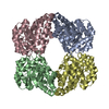
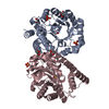
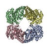
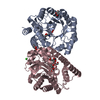
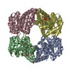
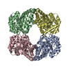
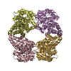
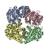
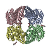
| Title | CRYSTAL STRUCTURE ANALYSIS OF N-ACETYLNEURAMINATE LYASE FROM HAEMOPHILUS INFLUENZAE: CRYSTAL FORM II IN COMPLEX WITH 4-OXO-SIALIC ACID | ||||||
 Components Components | N-ACETYL-NEURAMINATE LYASE | ||||||
 Keywords Keywords | LYASE / BETA BARREL | ||||||
| Function / homology |  Function and homology information Function and homology informationN-acetylneuraminate lyase / N-acetylneuraminate lyase activity / N-acetylneuraminate catabolic process / carbohydrate metabolic process / cytosol Similarity search - Function | ||||||
| Biological species |  Haemophilus influenzae (bacteria) Haemophilus influenzae (bacteria) | ||||||
| Method |  X-RAY DIFFRACTION / Resolution: 1.8 Å X-RAY DIFFRACTION / Resolution: 1.8 Å | ||||||
 Authors Authors | Barbosa, J.A.R.G. / Smith, B.J. / DeGori, R. / Lawrence, M.C. | ||||||
 Citation Citation | Journal: J.Mol.Biol. / Year: 2000 Title: Active site modulation in the N-acetylneuraminate lyase sub-family as revealed by the structure of the inhibitor-complexed Haemophilus influenzae enzyme. Authors: Barbosa, J.A.R.G. / Smith, B.J. / DeGori, R. / Ooi, H.C. / Marcuccio, S.M. / Campi, E.M. / Jackson, W.R. / Brossmer, R. / Sommer, M. / Lawrence, M.C. | ||||||
| History |
|
- Structure visualization
| Structure viewer | Molecule: MolmilJmol/JSmol |
|---|
- Downloads & links
-Download
| PDBx/mmCIF format | 1f7b.cif.gz | 145.5 KB | Display | PDBx/mmCIF format |
|---|---|---|---|---|
| PDB format | pdb1f7b.ent.gz | 112 KB | Display | PDB format |
| PDBx/mmJSON format | 1f7b.json.gz | Tree view | PDBx/mmJSON format | |
| Others |  Other downloads Other downloads |
-Validation report
| Arichive directory | https://data.pdbj.org/pub/pdb/validation_reports/f7/1f7bftp://data.pdbj.org/pub/pdb/validation_reports/f7/1f7b | HTTPS FTP |
|---|
-Related structure data


-Links
 PDBj
PDBj- Assembly
| Deposited unit | 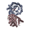
| ||||||||
|---|---|---|---|---|---|---|---|---|---|
| 1 | 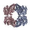
| ||||||||
| Unit cell |
| ||||||||
| Details | The biological assembly is a tetramer constructed from chains A and C generated by the two-fold. |
-Components
| #1: Protein | Mass: 32561.570 Da / Num. of mol.: 2 Source method: isolated from a genetically manipulated source Source: (gene. exp.) Haemophilus influenzae (bacteria) / Plasmid: PKKTAC / Production host: #2: Chemical | ChemComp-NAU / |   Mass: 311.286 Da / Num. of mol.: 1 / Source method: obtained synthetically / Formula: C11H21NO9 Mass: 311.286 Da / Num. of mol.: 1 / Source method: obtained synthetically / Formula: C11H21NO9#3: Chemical | ChemComp-CL / |   Mass: 35.453 Da / Num. of mol.: 1 / Source method: obtained synthetically / Formula: Cl Mass: 35.453 Da / Num. of mol.: 1 / Source method: obtained synthetically / Formula: Cl#4: Chemical | ChemComp-NAV / |   Mass: 293.270 Da / Num. of mol.: 1 / Source method: obtained synthetically / Formula: C11H19NO8 Mass: 293.270 Da / Num. of mol.: 1 / Source method: obtained synthetically / Formula: C11H19NO8#5: Water | ChemComp-HOH / |  Mass: 18.015 Da / Num. of mol.: 792 / Source method: isolated from a natural source / Formula: H2O Mass: 18.015 Da / Num. of mol.: 792 / Source method: isolated from a natural source / Formula: H2OHas protein modification | Y | |
|---|
-Experimental details
-Experiment
| Experiment | Method: X-RAY DIFFRACTION / Number of used crystals: 1 |
|---|
- Sample preparation
| Crystal | Density Matthews: 2.4 Å3/Da / Density % sol: 48.84 % | ||||||||||||||||||||||||||||||||||||||||||
|---|---|---|---|---|---|---|---|---|---|---|---|---|---|---|---|---|---|---|---|---|---|---|---|---|---|---|---|---|---|---|---|---|---|---|---|---|---|---|---|---|---|---|---|
| Crystal grow | Temperature: 293 K / Method: vapor diffusion, hanging drop / pH: 4.6 Details: PEG 4000, sodium acetate, ammonium acetate, pH 4.6, VAPOR DIFFUSION, HANGING DROP, temperature 293K | ||||||||||||||||||||||||||||||||||||||||||
| Crystal grow | *PLUS pH: 7.5 | ||||||||||||||||||||||||||||||||||||||||||
| Components of the solutions | *PLUS
|
-Data collection
| Diffraction | Mean temperature: 113 K |
|---|---|
| Diffraction source | Source: ROTATING ANODE / Type: MACSCIENCE / Wavelength: 1.5418 |
| Detector | Type: RIGAKU RAXIS II / Detector: IMAGE PLATE |
| Radiation | Protocol: SINGLE WAVELENGTH / Monochromatic (M) / Laue (L): M / Scattering type: x-ray |
| Radiation wavelength | Wavelength: 1.5418 Å / Relative weight: 1 |
| Reflection | Resolution: 1.8→15 Å / Num. all: 59168 / Num. obs: 52684 / % possible obs: 89 % / Observed criterion σ(F): 0 / Observed criterion σ(I): 0 / Redundancy: 4.4 % / Rmerge(I) obs: 0.06 / Net I/σ(I): 21 |
| Reflection shell | Resolution: 1.8→1.86 Å / Redundancy: 2.7 % / Rmerge(I) obs: 0.2 / Num. unique all: 5816 / % possible all: 42.4 |
| Reflection | *PLUS Num. measured all: 231047 |
| Reflection shell | *PLUS % possible obs: 42.4 % / Rmerge(I) obs: 0.2 / Mean I/σ(I) obs: 5.2 |
- Processing
| Software |
| |||||||||||||||||||||||||
|---|---|---|---|---|---|---|---|---|---|---|---|---|---|---|---|---|---|---|---|---|---|---|---|---|---|---|
| Refinement | Resolution: 1.8→15 Å / σ(F): 0 / σ(I): 0 / Stereochemistry target values: Engh & Huber
| |||||||||||||||||||||||||
| Refinement step | Cycle: LAST / Resolution: 1.8→15 Å
| |||||||||||||||||||||||||
| Refine LS restraints |
| |||||||||||||||||||||||||
| Software | *PLUS Name: REFMAC / Classification: refinement | |||||||||||||||||||||||||
| Refinement | *PLUS σ(F): 0 / Rfactor obs: 0.167 / Rfactor Rfree: 0.21 | |||||||||||||||||||||||||
| Solvent computation | *PLUS | |||||||||||||||||||||||||
| Displacement parameters | *PLUS | |||||||||||||||||||||||||
| Refine LS restraints | *PLUS
|
