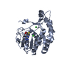
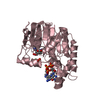
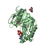
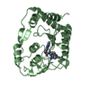
Entry Database : PDB / ID : 5kqsTitle Structure of NS5 methyltransferase from Zika virus bound to S-adenosylmethionine and 7-methyl-guanosine-5'-diphosphate Methyltransferase Keywords / / / / Function / homology Function Domain/homology Component
/ / / / / / / / / / / / / / / / / / / / / / / / / / / / / / / / / / / / / / / / / / / / / / / / / / / / / / / / / / / / / / / / / / / / / / / / / / / / / / / / / / / / / / / / / / / / / / / / / / / / / / / / / / / / / / / / / / / / / / / / / / / / / / / / / / / / Biological species Method / / / / Resolution : 1.5 Å Authors Coloma, J. / Jain, R. / Rajashankar, K.R. / Aggarwal, A.K. Journal : Cell Rep / Year : 2016Title : Structures of NS5 Methyltransferase from Zika Virus.Authors : Coloma, J. / Jain, R. / Rajashankar, K.R. / Garcia-Sastre, A. / Aggarwal, A.K. History Deposition Jul 6, 2016 Deposition site / Processing site Revision 1.0 Sep 14, 2016 Provider / Type Revision 1.1 Jan 4, 2017 Group Revision 1.2 Aug 3, 2022 Group / Structure summary / Category / entityItem / _database_2.pdbx_database_accession / _entity.pdbx_descriptionRevision 1.3 Oct 4, 2023 Group / Refinement descriptionCategory / chem_comp_bond / pdbx_initial_refinement_model
Show all Show less
 Movie
Movie Controller

 Yorodumi
 Open data
 Basic information
 Components
 Keywords
 Function and homology information
 Zika virus
 X-RAY DIFFRACTION /
  Authors
 Citation
 Structure visualization
 Downloads & links
 Other downloads
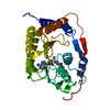


 PDBj
PDBj


 Assembly
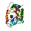


 Mass: 459.243 Da / Num. of mol.: 1 / Source method: obtained synthetically / Formula: C11H19N5O11P2
 Mass: 92.094 Da / Num. of mol.: 4 / Source method: obtained synthetically / Formula: C3H8O3
 Mass: 59.044 Da / Num. of mol.: 1 / Source method: obtained synthetically / Formula: C2H3O2
 Mass: 398.437 Da / Num. of mol.: 1 / Source method: obtained synthetically / Formula: C15H22N6O5S
 Mass: 94.971 Da / Num. of mol.: 1 / Source method: obtained synthetically / Formula: PO4
 Sample preparation
 / Beamline: 23-ID-D / Wavelength: 1.0332 Å
 Processing