 Movie
Movie Controller


+ Open data

- Basic information
| Entry | Database: PDB / ID: 5ko4 | ||||||
|---|---|---|---|---|---|---|---|
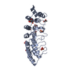
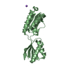
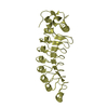
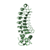
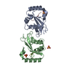
| Title | Bromodomain from Trypanosoma brucei Tb427.10.8150 | ||||||
 Components Components | Putative uncharacterized protein | ||||||
 Keywords Keywords | UNKNOWN FUNCTION / BROMODOMAIN / Structural Genomics Consortium (SGC) | ||||||
| Function / homology |  Function and homology information Function and homology information | ||||||
| Biological species |  | ||||||
| Method |  X-RAY DIFFRACTION / SYNCHROTRON / MOLECULAR REPLACEMENT / molecular replacement / Resolution: 1.44 Å X-RAY DIFFRACTION / SYNCHROTRON / MOLECULAR REPLACEMENT / molecular replacement / Resolution: 1.44 Å | ||||||
 Authors Authors | El Bakkouri, M. / Walker, J.R. / Hou, C.F.D. / Lin, Y.H. / Bountra, C. / Edwards, A.M. / Arrowsmith, C.H. / Hui, R. / Structural Genomics Consortium (SGC) | ||||||
 Citation Citation | Journal: To be published Title: Bromodomain from Trypanosoma brucei Tb427.10.8150 Authors: El Bakkouri, M. / Walker, J.R. / Hou, C.F.D. / Lin, Y.H. / Bountra, C. / Edwards, A.M. / Arrowsmith, C.H. / Hui, R. / Structural Genomics Consortium (SGC) | ||||||
| History |
|
- Structure visualization
| Structure viewer | Molecule: MolmilJmol/JSmol |
|---|
- Downloads & links
-Download
| PDBx/mmCIF format | 5ko4.cif.gz | 111.8 KB | Display | PDBx/mmCIF format |
|---|---|---|---|---|
| PDB format | pdb5ko4.ent.gz | 87.7 KB | Display | PDB format |
| PDBx/mmJSON format | 5ko4.json.gz | Tree view | PDBx/mmJSON format | |
| Others |  Other downloads Other downloads |
-Validation report
| Arichive directory | https://data.pdbj.org/pub/pdb/validation_reports/ko/5ko4ftp://data.pdbj.org/pub/pdb/validation_reports/ko/5ko4 | HTTPS FTP |
|---|
-Related structure data
| Related structure data |  3o33S S: Starting model for refinement |
|---|---|
| Similar structure data |


-Links
 PDBj
PDBj- Assembly
| Deposited unit | 
| ||||||||
|---|---|---|---|---|---|---|---|---|---|
| 1 |
| ||||||||
| Unit cell |
|
-Components
| #1: Protein | Mass: 12780.618 Da / Num. of mol.: 2 / Fragment: UNP residues 25-130 Source method: isolated from a genetically manipulated source Source: (gene. exp.) Strain: 927/4 GUTat10.1 / Gene: Tb10.6k15.2370 / Plasmid: PET15-MHL / Production host:  #2: Chemical |   Mass: 96.063 Da / Num. of mol.: 3 / Source method: obtained synthetically / Formula: SO4 Mass: 96.063 Da / Num. of mol.: 3 / Source method: obtained synthetically / Formula: SO4#3: Water | ChemComp-HOH / |  Mass: 18.015 Da / Num. of mol.: 255 / Source method: isolated from a natural source / Formula: H2O Mass: 18.015 Da / Num. of mol.: 255 / Source method: isolated from a natural source / Formula: H2O |
|---|
-Experimental details
-Experiment
| Experiment | Method: X-RAY DIFFRACTION / Number of used crystals: 1 |
|---|
- Sample preparation
| Crystal | Density Matthews: 2.06 Å3/Da / Density % sol: 40.39 % / Mosaicity: 0.42 ° |
|---|---|
| Crystal grow | Temperature: 293 K / Method: vapor diffusion, sitting drop / pH: 6.5 Details: The 12 mg/ml (0.95 mM) protein in the base buffer (100 mM NaCl, 0.5 mM TCEP, 20 mM HEPES pH7,5) was crystallized at 20 ??C in 30% PEGM5K, 0.2 M NH4SO4, 0.1 M MES pH 6.5, in vapor diffusion ...Details: The 12 mg/ml (0.95 mM) protein in the base buffer (100 mM NaCl, 0.5 mM TCEP, 20 mM HEPES pH7,5) was crystallized at 20 ??C in 30% PEGM5K, 0.2 M NH4SO4, 0.1 M MES pH 6.5, in vapor diffusion sitting drop method. Final concentration of 3% DMSO and 1 mM SGC-CBP30 (IUPAC name: 8-(3-chloro-4-methoxy-phenethyl)-4-(3,5-dimethyl-isoxazol-4-yl)-9-(2-(morpholin-4-yl)-propyl)-7,9-diaza-bicyclo[4.3.0]nona-1(6),2,4,7-tetraene) was added to the concentrated protein immediately prior to setting up the crystallization plate. |
-Data collection
| Diffraction | Mean temperature: 100 K |
|---|---|
| Diffraction source | Source: SYNCHROTRON / Site: APS  / Beamline: 22-ID / Wavelength: 0.97901 Å / Beamline: 22-ID / Wavelength: 0.97901 Å |
| Detector | Type: MARMOSAIC 300 mm CCD / Detector: CCD / Date: Apr 3, 2016 |
| Radiation | Protocol: SINGLE WAVELENGTH / Monochromatic (M) / Laue (L): M / Scattering type: x-ray |
| Radiation wavelength | Wavelength: 0.97901 Å / Relative weight: 1 |
| Reflection | Resolution: 1.44→50 Å / Num. obs: 35635 / % possible obs: 99.2 % / Redundancy: 6.7 % / Biso Wilson estimate: 15.47 Å2 / Rmerge(I) obs: 0.082 / Net I/σ(I): 9.1 |
| Reflection shell | Resolution: 1.44→1.46 Å / Redundancy: 4.5 % / Rmerge(I) obs: 0.358 / % possible all: 96.5 |
-Phasing
| Phasing | Method: molecular replacement |
|---|
- Processing
| Software |
| ||||||||||||||||||||||||||||||||||||||||||||||||||||||||||||||||||||||||||||||||||||||||||||||||||||||||||||||||||
|---|---|---|---|---|---|---|---|---|---|---|---|---|---|---|---|---|---|---|---|---|---|---|---|---|---|---|---|---|---|---|---|---|---|---|---|---|---|---|---|---|---|---|---|---|---|---|---|---|---|---|---|---|---|---|---|---|---|---|---|---|---|---|---|---|---|---|---|---|---|---|---|---|---|---|---|---|---|---|---|---|---|---|---|---|---|---|---|---|---|---|---|---|---|---|---|---|---|---|---|---|---|---|---|---|---|---|---|---|---|---|---|---|---|---|---|
| Refinement | Method to determine structure: MOLECULAR REPLACEMENT Starting model: 3O33 Resolution: 1.44→41.8 Å / Cor.coef. Fo:Fc: 0.964 / Cor.coef. Fo:Fc free: 0.956 / Rfactor Rfree error: 0 / SU R Cruickshank DPI: 0.076 / Cross valid method: THROUGHOUT / σ(F): 0 / SU R Blow DPI: 0.069 / SU Rfree Blow DPI: 0.065 / SU Rfree Cruickshank DPI: 0.062
| ||||||||||||||||||||||||||||||||||||||||||||||||||||||||||||||||||||||||||||||||||||||||||||||||||||||||||||||||||
| Displacement parameters | Biso mean: 17.65 Å2
| ||||||||||||||||||||||||||||||||||||||||||||||||||||||||||||||||||||||||||||||||||||||||||||||||||||||||||||||||||
| Refine analyze | Luzzati coordinate error obs: 0.15 Å | ||||||||||||||||||||||||||||||||||||||||||||||||||||||||||||||||||||||||||||||||||||||||||||||||||||||||||||||||||
| Refinement step | Cycle: LAST / Resolution: 1.44→41.8 Å
| ||||||||||||||||||||||||||||||||||||||||||||||||||||||||||||||||||||||||||||||||||||||||||||||||||||||||||||||||||
| Refine LS restraints |
| ||||||||||||||||||||||||||||||||||||||||||||||||||||||||||||||||||||||||||||||||||||||||||||||||||||||||||||||||||
| LS refinement shell | Resolution: 1.44→1.48 Å / Rfactor Rfree error: 0 / Total num. of bins used: 18
| ||||||||||||||||||||||||||||||||||||||||||||||||||||||||||||||||||||||||||||||||||||||||||||||||||||||||||||||||||
| Refinement TLS params. | Method: refined / Refine-ID: X-RAY DIFFRACTION
| ||||||||||||||||||||||||||||||||||||||||||||||||||||||||||||||||||||||||||||||||||||||||||||||||||||||||||||||||||
| Refinement TLS group |
|
