 Movie
Movie Controller


+ Open data

- Basic information
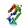
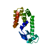
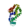
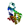
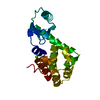
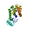
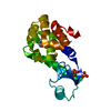
| Entry | Database: PDB / ID: 5khz | ||||||
|---|---|---|---|---|---|---|---|
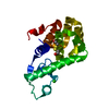
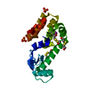
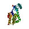
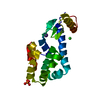
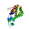
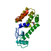
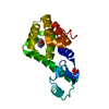
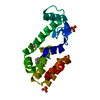
| Title | PSEUDO T4 LYSOZYME | ||||||
 Components Components | Endolysin | ||||||
 Keywords Keywords | HYDROLASE / HALOGEN BONDS / T4 LYSOZYME | ||||||
| Function / homology |  Function and homology information Function and homology informationviral release from host cell by cytolysis / peptidoglycan catabolic process / cell wall macromolecule catabolic process / lysozyme / lysozyme activity / host cell cytoplasm / defense response to bacterium Similarity search - Function | ||||||
| Biological species |  Enterobacteria phage T4 (virus) Enterobacteria phage T4 (virus) | ||||||
| Method |  X-RAY DIFFRACTION / MOLECULAR REPLACEMENT / Resolution: 1.49 Å X-RAY DIFFRACTION / MOLECULAR REPLACEMENT / Resolution: 1.49 Å | ||||||
 Authors Authors | Scholfield, M.R. | ||||||
| Funding support |  United States, 1items United States, 1items
| ||||||
 Citation Citation | Journal: Biochemistry / Year: 2017 Title: Structure-Energy Relationships of Halogen Bonds in Proteins. Authors: Scholfield, M.R. / Ford, M.C. / Carlsson, A.C. / Butta, H. / Mehl, R.A. / Ho, P.S. | ||||||
| History |
|
- Structure visualization
| Structure viewer | Molecule: MolmilJmol/JSmol |
|---|
- Downloads & links
-Download
| PDBx/mmCIF format | 5khz.cif.gz | 89.2 KB | Display | PDBx/mmCIF format |
|---|---|---|---|---|
| PDB format | pdb5khz.ent.gz | 66.3 KB | Display | PDB format |
| PDBx/mmJSON format | 5khz.json.gz | Tree view | PDBx/mmJSON format | |
| Others |  Other downloads Other downloads |
-Validation report
| Arichive directory | https://data.pdbj.org/pub/pdb/validation_reports/kh/5khzftp://data.pdbj.org/pub/pdb/validation_reports/kh/5khz | HTTPS FTP |
|---|
-Related structure data
| Related structure data | 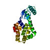 5ki1C 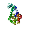 5ki2C 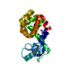 5ki3C 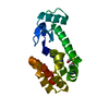 5ki8C 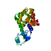 5kigC 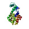 5kiiC 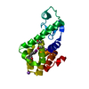 5kimC 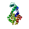 5kioC 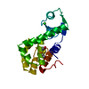 1l63S C: citing same article ( S: Starting model for refinement |
|---|---|
| Similar structure data |


-Links
 PDBj
PDBj


- Assembly
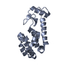
| Deposited unit | 
| ||||||||||||
|---|---|---|---|---|---|---|---|---|---|---|---|---|---|
| 1 |
| ||||||||||||
| Unit cell |
| ||||||||||||
| Components on special symmetry positions |
|
-Components
| #1: Protein | Mass: 19457.246 Da / Num. of mol.: 1 / Mutation: YES Source method: isolated from a genetically manipulated source Source: (gene. exp.) Enterobacteria phage T4 (virus) / Plasmid: PBAD / Production host:  |
|---|---|
| #2: Chemical | ChemComp-HED /   Mass: 154.251 Da / Num. of mol.: 1 / Source method: obtained synthetically / Formula: C4H10O2S2 Mass: 154.251 Da / Num. of mol.: 1 / Source method: obtained synthetically / Formula: C4H10O2S2 |
| #3: Water | ChemComp-HOH /  Mass: 18.015 Da / Num. of mol.: 375 / Source method: isolated from a natural source / Formula: H2O Mass: 18.015 Da / Num. of mol.: 375 / Source method: isolated from a natural source / Formula: H2O |
-Experimental details
-Experiment
| Experiment | Method: X-RAY DIFFRACTION / Number of used crystals: 1 |
|---|
- Sample preparation
| Crystal | Density Matthews: 2.75 Å3/Da / Density % sol: 55.21 % |
|---|---|
| Crystal grow | Temperature: 291.15 K / Method: vapor diffusion, hanging drop / pH: 6.9 Details: 2.0M KPO4, 50MM 2-HYDROXYETHYLDISULFIDE, 50MM 2-MERCAPTOETHANOL, PH 6.9 PH range: 6.9 |
-Data collection
| Diffraction | Mean temperature: 100 K |
|---|---|
| Diffraction source | Source: ROTATING ANODE / Type: RIGAKU MICROMAX-003 / Wavelength: 1.54 Å |
| Detector | Type: DECTRIS PILATUS 200K / Detector: PIXEL / Date: Jun 13, 2014 |
| Radiation | Protocol: SINGLE WAVELENGTH / Monochromatic (M) / Laue (L): M / Scattering type: x-ray |
| Radiation wavelength | Wavelength: 1.54 Å / Relative weight: 1 |
| Reflection | Resolution: 1.49→21.98 Å / Num. obs: 31370 / % possible obs: 92 % / Redundancy: 9.5 % / Biso Wilson estimate: 11.77 Å2 / Rmerge(I) obs: 0.085 / Rsym value: 0.089 / Net I/σ(I): 28.4 |
- Processing
| Software |
| |||||||||||||||||||||||||||||||||||||||||||||||||||||||||||||||||||||||||||||||||||||||||||||||||||||||||
|---|---|---|---|---|---|---|---|---|---|---|---|---|---|---|---|---|---|---|---|---|---|---|---|---|---|---|---|---|---|---|---|---|---|---|---|---|---|---|---|---|---|---|---|---|---|---|---|---|---|---|---|---|---|---|---|---|---|---|---|---|---|---|---|---|---|---|---|---|---|---|---|---|---|---|---|---|---|---|---|---|---|---|---|---|---|---|---|---|---|---|---|---|---|---|---|---|---|---|---|---|---|---|---|---|---|---|
| Refinement | Method to determine structure: MOLECULAR REPLACEMENT Starting model: 1L63 Resolution: 1.49→21.98 Å / SU ML: 0.133 / Cross valid method: FREE R-VALUE / σ(F): 1.33 / Phase error: 18.276
| |||||||||||||||||||||||||||||||||||||||||||||||||||||||||||||||||||||||||||||||||||||||||||||||||||||||||
| Solvent computation | Shrinkage radii: 0.9 Å / VDW probe radii: 1.11 Å | |||||||||||||||||||||||||||||||||||||||||||||||||||||||||||||||||||||||||||||||||||||||||||||||||||||||||
| Displacement parameters | Biso mean: 17.05 Å2 | |||||||||||||||||||||||||||||||||||||||||||||||||||||||||||||||||||||||||||||||||||||||||||||||||||||||||
| Refinement step | Cycle: LAST / Resolution: 1.49→21.98 Å
| |||||||||||||||||||||||||||||||||||||||||||||||||||||||||||||||||||||||||||||||||||||||||||||||||||||||||
| Refine LS restraints |
| |||||||||||||||||||||||||||||||||||||||||||||||||||||||||||||||||||||||||||||||||||||||||||||||||||||||||
| LS refinement shell |
|
