[English] 日本語
 Yorodumi
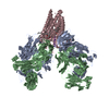
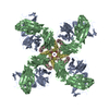
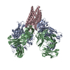
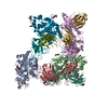
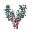
- PDB-5ken: EBOV GP in complex with variable Fab domains of IgGs c4G7 and c13C6 -
+ Open data
- Basic information
| Entry | Database: PDB / ID: 5ken | ||||||||||||
|---|---|---|---|---|---|---|---|---|---|---|---|---|---|
| Title | EBOV GP in complex with variable Fab domains of IgGs c4G7 and c13C6 | ||||||||||||
 Components Components |
| ||||||||||||
 Keywords Keywords | VIRAL PROTEIN/IMMUNE SYSTEM / Ebola virus surface glycoprotein / therapeutic antibody cocktail / ZMapp / VIRAL PROTEIN-IMMUNE SYSTEM complex | ||||||||||||
| Function / homology |  Function and homology information Function and homology informationsymbiont-mediated killing of host cell / host cell endoplasmic reticulum / viral budding from plasma membrane / clathrin-dependent endocytosis of virus by host cell / symbiont-mediated-mediated suppression of host tetherin activity / entry receptor-mediated virion attachment to host cell / host cell cytoplasm / symbiont-mediated suppression of host innate immune response / membrane raft / fusion of virus membrane with host endosome membrane ...symbiont-mediated killing of host cell / host cell endoplasmic reticulum / viral budding from plasma membrane / clathrin-dependent endocytosis of virus by host cell / symbiont-mediated-mediated suppression of host tetherin activity / entry receptor-mediated virion attachment to host cell / host cell cytoplasm / symbiont-mediated suppression of host innate immune response / membrane raft / fusion of virus membrane with host endosome membrane / viral envelope / symbiont entry into host cell / lipid binding / host cell plasma membrane / virion membrane / extracellular region / identical protein binding Similarity search - Function | ||||||||||||
| Biological species |   Homo sapiens (human) Homo sapiens (human) | ||||||||||||
| Method | ELECTRON MICROSCOPY / single particle reconstruction / cryo EM / Resolution: 4.3 Å | ||||||||||||
 Authors Authors | Pallesen, J. / Murin, C.D. / de Val, N. / Cottrell, C.A. / Hastie, K.M. / Turner, H.L. / Fusco, M.L. / Flyak, A.I. / Zeitlin, L. / Crowe Jr., J.E. ...Pallesen, J. / Murin, C.D. / de Val, N. / Cottrell, C.A. / Hastie, K.M. / Turner, H.L. / Fusco, M.L. / Flyak, A.I. / Zeitlin, L. / Crowe Jr., J.E. / Andersen, K.G. / Saphire, E.O. / Ward, A.B. | ||||||||||||
| Funding support |  United States, 3items United States, 3items
| ||||||||||||
 Citation Citation |  Journal: Nat Microbiol / Year: 2016 Journal: Nat Microbiol / Year: 2016Title: Structures of Ebola virus GP and sGP in complex with therapeutic antibodies. Authors: Jesper Pallesen / Charles D Murin / Natalia de Val / Christopher A Cottrell / Kathryn M Hastie / Hannah L Turner / Marnie L Fusco / Andrew I Flyak / Larry Zeitlin / James E Crowe / Kristian ...Authors: Jesper Pallesen / Charles D Murin / Natalia de Val / Christopher A Cottrell / Kathryn M Hastie / Hannah L Turner / Marnie L Fusco / Andrew I Flyak / Larry Zeitlin / James E Crowe / Kristian G Andersen / Erica Ollmann Saphire / Andrew B Ward /  Abstract: The Ebola virus (EBOV) GP gene encodes two glycoproteins. The major product is a soluble, dimeric glycoprotein (sGP) that is secreted abundantly. Despite the abundance of sGP during infection, little ...The Ebola virus (EBOV) GP gene encodes two glycoproteins. The major product is a soluble, dimeric glycoprotein (sGP) that is secreted abundantly. Despite the abundance of sGP during infection, little is known regarding its structure or functional role. A minor product, resulting from transcriptional editing, is the transmembrane-anchored, trimeric viral surface glycoprotein (GP). GP mediates attachment to and entry into host cells, and is the intended target of antibody therapeutics. Because large portions of sequence are shared between GP and sGP, it has been hypothesized that sGP may potentially subvert the immune response or may contribute to pathogenicity. In this study, we present cryo-electron microscopy structures of GP and sGP in complex with GP-specific and GP/sGP cross-reactive antibodies undergoing human clinical trials. The structure of the sGP dimer presented here, in complex with both an sGP-specific antibody and a GP/sGP cross-reactive antibody, permits us to unambiguously assign the oligomeric arrangement of sGP and compare its structure and epitope presentation to those of GP. We also provide biophysical evaluation of naturally occurring GP/sGP mutations that fall within the footprints identified by our high-resolution structures. Taken together, our data provide a detailed and more complete picture of the accessible Ebolavirus glycoprotein landscape and a structural basis to evaluate patient and vaccine antibody responses towards differently structured products of the GP gene. | ||||||||||||
| History |
|
- Structure visualization
| Movie |
 Movie viewer Movie viewer |
|---|---|
| Structure viewer | Molecule:  Molmil Molmil Jmol/JSmol Jmol/JSmol |
- Downloads & links
- Download
| PDBx/mmCIF format |  5ken.cif.gz 5ken.cif.gz | 427.7 KB | Display |  PDBx/mmCIF format PDBx/mmCIF format |
|---|---|---|---|---|
| PDB format |  pdb5ken.ent.gz pdb5ken.ent.gz | 346.9 KB | Display |  PDB format PDB format |
| PDBx/mmJSON format |  5ken.json.gz 5ken.json.gz | Tree view |  PDBx/mmJSON format PDBx/mmJSON format | |
| Others |  Other downloads Other downloads |
-Validation report
| Arichive directory |  https://data.pdbj.org/pub/pdb/validation_reports/ke/5ken https://data.pdbj.org/pub/pdb/validation_reports/ke/5ken ftp://data.pdbj.org/pub/pdb/validation_reports/ke/5ken ftp://data.pdbj.org/pub/pdb/validation_reports/ke/5ken | HTTPS FTP |
|---|
-Related structure data
| Related structure data |  8242MC  8240C  8241C  5kelC  5kemC M: map data used to model this data C: citing same article ( |
|---|---|
| Similar structure data |
- Links
Links
- Assembly
| Deposited unit | 
|
|---|---|
| 1 |
|
- Components
-Ebola surface glycoprotein, ... , 2 types, 6 molecules AEKBFM
| #1: Protein | Mass: 30910.719 Da / Num. of mol.: 3 Source method: isolated from a genetically manipulated source Source: (gene. exp.)   #2: Protein | Mass: 12650.368 Da / Num. of mol.: 3 Source method: isolated from a genetically manipulated source Source: (gene. exp.)   |
|---|
-Antibody , 4 types, 10 molecules CGNDHOIPJQ
| #3: Antibody | Mass: 12851.230 Da / Num. of mol.: 3 Source method: isolated from a genetically manipulated source Source: (gene. exp.)  Homo sapiens (human) / Production host: Homo sapiens (human) / Production host:  #4: Antibody | Mass: 11733.013 Da / Num. of mol.: 3 Source method: isolated from a genetically manipulated source Source: (gene. exp.)  Homo sapiens (human) / Production host: Homo sapiens (human) / Production host:  #5: Antibody | Mass: 11611.877 Da / Num. of mol.: 2 Source method: isolated from a genetically manipulated source Source: (gene. exp.)  Homo sapiens (human) / Production host: Homo sapiens (human) / Production host:  #6: Antibody | Mass: 13169.679 Da / Num. of mol.: 2 Source method: isolated from a genetically manipulated source Source: (gene. exp.)  Homo sapiens (human) / Production host: Homo sapiens (human) / Production host:  |
|---|
-Sugars , 3 types, 9 molecules 
| #7: Polysaccharide | Source method: isolated from a genetically manipulated source #8: Polysaccharide | Source method: isolated from a genetically manipulated source #9: Sugar | |
|---|
-Details
| Has protein modification | Y |
|---|
-Experimental details
-Experiment
| Experiment | Method: ELECTRON MICROSCOPY |
|---|---|
| EM experiment | Aggregation state: PARTICLE / 3D reconstruction method: single particle reconstruction |
- Sample preparation
| Component | Name: Ebola virus trimeric surface glycoprotein in complex with IgG c4G7 and c13C6 Fabs Type: COMPLEX / Entity ID: #1-#6 / Source: MULTIPLE SOURCES |
|---|---|
| Molecular weight | Value: 0.54 MDa / Experimental value: YES |
| Buffer solution | pH: 7.4 |
| Buffer component | Name: 1xTBS |
| Specimen | Conc.: 4 mg/ml / Embedding applied: NO / Shadowing applied: NO / Staining applied: NO / Vitrification applied: YES |
| Specimen support | Grid material: COPPER / Grid mesh size: 400 divisions/in. / Grid type: C-flat-2/2 4C |
| Vitrification | Instrument: HOMEMADE PLUNGER / Cryogen name: ETHANE / Chamber temperature: 277 K |
- Electron microscopy imaging
| Experimental equipment |  Model: Titan Krios / Image courtesy: FEI Company |
|---|---|
| Microscopy | Model: FEI TITAN KRIOS |
| Electron gun | Electron source:  FIELD EMISSION GUN / Accelerating voltage: 300 kV / Illumination mode: FLOOD BEAM FIELD EMISSION GUN / Accelerating voltage: 300 kV / Illumination mode: FLOOD BEAM |
| Electron lens | Mode: BRIGHT FIELD / Nominal magnification: 22500 X / Nominal defocus max: 4000 nm / Nominal defocus min: 1000 nm / Cs: 2.7 mm / Alignment procedure: COMA FREE |
| Specimen holder | Cryogen: NITROGEN / Specimen holder model: FEI TITAN KRIOS AUTOGRID HOLDER / Temperature (max): 77 K / Temperature (min): 77 K |
| Image recording | Average exposure time: 10 sec. / Electron dose: 57 e/Å2 / Detector mode: COUNTING / Film or detector model: GATAN K2 SUMMIT (4k x 4k) / Num. of grids imaged: 1 / Num. of real images: 2555 |
| Image scans | Movie frames/image: 50 / Used frames/image: 1-50 |
- Processing
| EM software |
| ||||||||||||||||||||||||||||||||||||||||||||||||||||
|---|---|---|---|---|---|---|---|---|---|---|---|---|---|---|---|---|---|---|---|---|---|---|---|---|---|---|---|---|---|---|---|---|---|---|---|---|---|---|---|---|---|---|---|---|---|---|---|---|---|---|---|---|---|
| CTF correction | Type: PHASE FLIPPING AND AMPLITUDE CORRECTION | ||||||||||||||||||||||||||||||||||||||||||||||||||||
| 3D reconstruction | Resolution: 4.3 Å / Resolution method: FSC 0.143 CUT-OFF / Num. of particles: 73000 / Num. of class averages: 6 / Symmetry type: POINT | ||||||||||||||||||||||||||||||||||||||||||||||||||||
| Atomic model building | Protocol: FLEXIBLE FIT / Space: REAL / Target criteria: EMRinger Details: Model building and refinement were conducted using a combination of software programs. The refinement target was optimizing using the MolProbity score while maintaining a high (good) EMRinger score. |
 Movie
Movie Controller



 PDBj
PDBj


