[English] 日本語
 Yorodumi
- PDB-5kbx: Co-crystal structure of the Saccharomyces cerevisiae histidine ph... -
+ Open data
- Basic information
| Entry | Database: PDB / ID: 5kbx | |||||||||
|---|---|---|---|---|---|---|---|---|---|---|
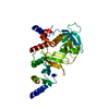
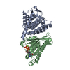
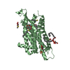
| Title | Co-crystal structure of the Saccharomyces cerevisiae histidine phosphotransfer signaling protein Ypd1 and the receiver domain of its downstream response regulator Ssk1 | |||||||||
 Components Components |
| |||||||||
 Keywords Keywords | SIGNALING PROTEIN / two-component signaling / phosphorelay / Ypd1 / Ssk1 / response regulator / histidine phosphotransfer protein / Saccharomyces cerevisiae / co-crystal / phosphotransfer | |||||||||
| Function / homology |  Function and homology information Function and homology informationtransferase activity, transferring phosphorus-containing groups / protein histidine kinase binding / histidine phosphotransfer kinase activity / regulation of p38MAPK cascade / osmosensory signaling via phosphorelay pathway / cellular hyperosmotic response / cellular response to acidic pH / mitogen-activated protein kinase kinase kinase binding / phosphorelay response regulator activity / phosphorelay signal transduction system ...transferase activity, transferring phosphorus-containing groups / protein histidine kinase binding / histidine phosphotransfer kinase activity / regulation of p38MAPK cascade / osmosensory signaling via phosphorelay pathway / cellular hyperosmotic response / cellular response to acidic pH / mitogen-activated protein kinase kinase kinase binding / phosphorelay response regulator activity / phosphorelay signal transduction system / protein kinase activator activity / regulation of actin cytoskeleton organization / nucleus / cytoplasm Similarity search - Function | |||||||||
| Biological species |  | |||||||||
| Method |  X-RAY DIFFRACTION / X-RAY DIFFRACTION /  SYNCHROTRON / SYNCHROTRON /  MAD / Resolution: 2.8 Å MAD / Resolution: 2.8 Å | |||||||||
 Authors Authors | Menon, S.K. / West, A.H. | |||||||||
| Funding support |  United States, 1items United States, 1items
| |||||||||
 Citation Citation |  Journal: To Be Published Journal: To Be PublishedTitle: Insights revealed by the co-crystal structure of the Saccharomyces cerevisiae histidine phosphotransfer signaling protein Ypd1 and the receiver domain of its downstream response regulator Ssk1 Authors: Menon, S.K. / Branscum, K.M. / Foster, C.A. / West, A.H. #1:  Journal: J. Mol. Biol. / Year: 2008 Journal: J. Mol. Biol. / Year: 2008Title: Crystal structure of a complex between the phosphorelay protein YPD1 and the response regulator domain of SLN1 bound to a phosphoryl analog. Authors: Zhao, X. / Copeland, D.M. / Soares, A.S. / West, A.H. #2: Journal: Acta Crystallogr. D Biol. Crystallogr. / Year: 2003 Title: Co-crystallization of the yeast phosphorelay protein YPD1 with the SLN1 response-regulator domain and preliminary X-ray diffraction analysis. Authors: Chooback, L. / West, A.H. | |||||||||
| History |
|
- Structure visualization
| Structure viewer | Molecule:  Molmil Molmil Jmol/JSmol Jmol/JSmol |
|---|
- Downloads & links
- Download
| PDBx/mmCIF format |  5kbx.cif.gz 5kbx.cif.gz | 164.3 KB | Display |  PDBx/mmCIF format PDBx/mmCIF format |
|---|---|---|---|---|
| PDB format |  pdb5kbx.ent.gz pdb5kbx.ent.gz | 107.7 KB | Display |  PDB format PDB format |
| PDBx/mmJSON format |  5kbx.json.gz 5kbx.json.gz | Tree view |  PDBx/mmJSON format PDBx/mmJSON format | |
| Others |  Other downloads Other downloads |
-Validation report
| Arichive directory |  https://data.pdbj.org/pub/pdb/validation_reports/kb/5kbx https://data.pdbj.org/pub/pdb/validation_reports/kb/5kbx ftp://data.pdbj.org/pub/pdb/validation_reports/kb/5kbx ftp://data.pdbj.org/pub/pdb/validation_reports/kb/5kbx | HTTPS FTP |
|---|
-Related structure data
| Similar structure data |
|---|
- Links
Links
- Assembly
| Deposited unit | 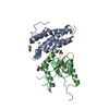
| ||||||||||
|---|---|---|---|---|---|---|---|---|---|---|---|
| 1 |
| ||||||||||
| Unit cell |
|
- Components
| #1: Protein | Mass: 19187.652 Da / Num. of mol.: 1 Source method: isolated from a genetically manipulated source Source: (gene. exp.)   | ||||||||
|---|---|---|---|---|---|---|---|---|---|
| #2: Protein | Mass: 24457.375 Da / Num. of mol.: 1 / Fragment: UNP residues 495-712 Source method: isolated from a genetically manipulated source Source: (gene. exp.)   | ||||||||
| #3: Chemical | | #4: Chemical | #5: Water | ChemComp-HOH / | Has ligand of interest | N | Has protein modification | Y | |
-Experimental details
-Experiment
| Experiment | Method:  X-RAY DIFFRACTION / Number of used crystals: 1 X-RAY DIFFRACTION / Number of used crystals: 1 |
|---|
- Sample preparation
| Crystal | Density Matthews: 2.51 Å3/Da / Density % sol: 51 % |
|---|---|
| Crystal grow | Temperature: 296 K / Method: vapor diffusion, hanging drop / pH: 10.5 Details: 0.2 M lithium sulfate, 0.1 M CAPS/NaOH pH 10.5, 1.2 M NaH2PO4/0.8 M K2HPO4 |
-Data collection
| Diffraction | Mean temperature: 100 K / Ambient temp details: Liquid N2 / Serial crystal experiment: N | ||||||||||||
|---|---|---|---|---|---|---|---|---|---|---|---|---|---|
| Diffraction source | Source:  SYNCHROTRON / Site: SYNCHROTRON / Site:  SSRL SSRL  / Beamline: BL11-1 / Wavelength: 0.97910, 0.97930, 0.9116 / Beamline: BL11-1 / Wavelength: 0.97910, 0.97930, 0.9116 | ||||||||||||
| Detector | Type: DECTRIS PILATUS 6M / Detector: PIXEL / Date: Sep 19, 2014 / Details: Rh coated flat | ||||||||||||
| Radiation | Monochromator: Si(111) Side scattering bent cube-root I-beam single crystal; asymmetric cut 4.965 degs Protocol: MAD / Monochromatic (M) / Laue (L): M / Scattering type: x-ray | ||||||||||||
| Radiation wavelength |
| ||||||||||||
| Reflection | Resolution: 2.798→33.7 Å / Num. obs: 10695 / % possible obs: 89 % / Observed criterion σ(I): -3 / Redundancy: 1.9 % / Biso Wilson estimate: 42.08 Å2 / CC1/2: 0.998 / Rmerge(I) obs: 0.037 / Net I/σ(I): 34 | ||||||||||||
| Reflection shell | Resolution: 2.8→2.9 Å / Redundancy: 1.8 % / Rmerge(I) obs: 0.168 / Mean I/σ(I) obs: 9.2 / Num. unique obs: 1163 / % possible all: 44 |
- Processing
| Software |
| ||||||||||||||||||||||||||||||||||||||||||||||||||||||||||||||||||||||||||||||||||||||||||||||||||||||||||||||||||||||||||||||||||||||||||||||||||||||
|---|---|---|---|---|---|---|---|---|---|---|---|---|---|---|---|---|---|---|---|---|---|---|---|---|---|---|---|---|---|---|---|---|---|---|---|---|---|---|---|---|---|---|---|---|---|---|---|---|---|---|---|---|---|---|---|---|---|---|---|---|---|---|---|---|---|---|---|---|---|---|---|---|---|---|---|---|---|---|---|---|---|---|---|---|---|---|---|---|---|---|---|---|---|---|---|---|---|---|---|---|---|---|---|---|---|---|---|---|---|---|---|---|---|---|---|---|---|---|---|---|---|---|---|---|---|---|---|---|---|---|---|---|---|---|---|---|---|---|---|---|---|---|---|---|---|---|---|---|---|---|---|
| Refinement | Method to determine structure:  MAD / Resolution: 2.8→33.27 Å / SU ML: 0.3645 / Cross valid method: FREE R-VALUE / σ(F): 1.33 / Phase error: 28.5826 MAD / Resolution: 2.8→33.27 Å / SU ML: 0.3645 / Cross valid method: FREE R-VALUE / σ(F): 1.33 / Phase error: 28.5826 Details: ellipsoidal truncation was applied to the data/ overall d_min = 2.8 d_min along a*= 3.1 d_min along b*= 3.1 d_min along c*= 2.8
| ||||||||||||||||||||||||||||||||||||||||||||||||||||||||||||||||||||||||||||||||||||||||||||||||||||||||||||||||||||||||||||||||||||||||||||||||||||||
| Solvent computation | Shrinkage radii: 0.9 Å / VDW probe radii: 1.11 Å | ||||||||||||||||||||||||||||||||||||||||||||||||||||||||||||||||||||||||||||||||||||||||||||||||||||||||||||||||||||||||||||||||||||||||||||||||||||||
| Displacement parameters | Biso mean: 49.18 Å2 | ||||||||||||||||||||||||||||||||||||||||||||||||||||||||||||||||||||||||||||||||||||||||||||||||||||||||||||||||||||||||||||||||||||||||||||||||||||||
| Refinement step | Cycle: LAST / Resolution: 2.8→33.27 Å
| ||||||||||||||||||||||||||||||||||||||||||||||||||||||||||||||||||||||||||||||||||||||||||||||||||||||||||||||||||||||||||||||||||||||||||||||||||||||
| Refine LS restraints |
| ||||||||||||||||||||||||||||||||||||||||||||||||||||||||||||||||||||||||||||||||||||||||||||||||||||||||||||||||||||||||||||||||||||||||||||||||||||||
| LS refinement shell |
| ||||||||||||||||||||||||||||||||||||||||||||||||||||||||||||||||||||||||||||||||||||||||||||||||||||||||||||||||||||||||||||||||||||||||||||||||||||||
| Refinement TLS params. | Method: refined / Refine-ID: X-RAY DIFFRACTION
| ||||||||||||||||||||||||||||||||||||||||||||||||||||||||||||||||||||||||||||||||||||||||||||||||||||||||||||||||||||||||||||||||||||||||||||||||||||||
| Refinement TLS group |
|
 Movie
Movie Controller





 PDBj
PDBj


