Monochromator: SI / Protocol: SINGLE WAVELENGTH / Monochromatic (M) / Laue (L): M / Scattering type: x-ray
Radiation wavelength
Wavelength: 0.95 Å / Relative weight: 1
Reflection
Resolution: 2.55→20 Å / Num. obs: 17367 / % possible obs: 99.5 % / Observed criterion σ(I): 2 / Redundancy: 3.79 % / Rmerge(I) obs: 0.1 / Net I/σ(I): 10.48
Reflection shell
Resolution: 2.55→2.6 Å / Redundancy: 3.86 % / Rmerge(I) obs: 0.57 / Mean I/σ(I) obs: 2.31 / % possible all: 97.5
-
Processing
Software
Name
Version
Classification
XDS
datareduction
Aimless
datascaling
Auto-Rickshaw
phasing
REFMAC
5.8.0103
refinement
Refinement
Method to determine structure: SAD / Resolution: 2.55→19.82 Å / Cor.coef. Fo:Fc: 0.948 / Cor.coef. Fo:Fc free: 0.898 / SU B: 12.824 / SU ML: 0.27 / Cross valid method: THROUGHOUT / ESU R: 0.519 / ESU R Free: 0.312 / Details: HYDROGENS HAVE BEEN ADDED IN THE RIDING POSITIONS
Rfactor
Num. reflection
% reflection
Selection details
Rfree
0.26708
1015
5.9 %
RANDOM
Rwork
0.19872
-
-
-
obs
0.20273
16280
99.29 %
-
Solvent computation
Ion probe radii: 0.8 Å / Shrinkage radii: 0.8 Å / VDW probe radii: 1.2 Å
 Movie
Movie Controller

 Yorodumi
 Open data
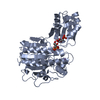
 Basic information
 Components
 Keywords
 Function and homology information
 Acinetobacter calcoaceticus (bacteria)
 X-RAY DIFFRACTION /
  Authors
 Citation
 Structure visualization
 Downloads & links
 Other downloads


 PDBj
PDBj
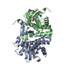
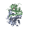
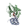
 Assembly

 Mass: 65.409 Da / Num. of mol.: 2 / Source method: obtained synthetically / Formula: Zn
 Mass: 128.556 Da / Num. of mol.: 2 / Source method: obtained synthetically / Formula: C6H5ClO
 Mass: 18.015 Da / Num. of mol.: 21 / Source method: isolated from a natural source / Formula: H2O
 Sample preparation
 / Beamline: MX2 / Wavelength: 0.95 Å
 Processing