Movie
Movie Controller

[English] 日本語
 Yorodumi
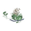
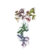
- PDB-5k1h: eIF3b relocated to the intersubunit face to interact with eIF1 an... -
+ Open data

- Basic information
| Entry | Database: PDB / ID: 5k1h | |||||||||
|---|---|---|---|---|---|---|---|---|---|---|
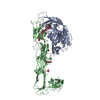
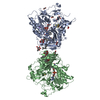
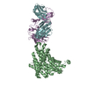
| Title | eIF3b relocated to the intersubunit face to interact with eIF1 and below the eIF2 ternary-complex. from the structure of a partial yeast 48S preinitiation complex in closed conformation. | |||||||||
 Components Components |
| |||||||||
 Keywords Keywords | TRANSLATION / eukaryotic translation initiation / ribosome / eIF3 peripheral subunits / cryo-EM | |||||||||
| Function / homology |  Function and homology information Function and homology informationviral translational termination-reinitiation / eukaryotic translation initiation factor 3 complex, eIF3m / IRES-dependent viral translational initiation / formation of cytoplasmic translation initiation complex / eukaryotic translation initiation factor 3 complex / eukaryotic 43S preinitiation complex / eukaryotic 48S preinitiation complex / regulation of translational initiation / Formation of the ternary complex, and subsequently, the 43S complex / Ribosomal scanning and start codon recognition ...viral translational termination-reinitiation / eukaryotic translation initiation factor 3 complex, eIF3m / IRES-dependent viral translational initiation / formation of cytoplasmic translation initiation complex / eukaryotic translation initiation factor 3 complex / eukaryotic 43S preinitiation complex / eukaryotic 48S preinitiation complex / regulation of translational initiation / Formation of the ternary complex, and subsequently, the 43S complex / Ribosomal scanning and start codon recognition / Translation initiation complex formation / Formation of a pool of free 40S subunits / GTP hydrolysis and joining of the 60S ribosomal subunit / L13a-mediated translational silencing of Ceruloplasmin expression / translation initiation factor activity / translation initiation factor binding / translational initiation / cytoplasmic stress granule / molecular adaptor activity / synapse / RNA binding / extracellular exosome / cytosol Similarity search - Function | |||||||||
| Biological species |  Homo sapiens (human) Homo sapiens (human) | |||||||||
| Method | ELECTRON MICROSCOPY / single particle reconstruction / cryo EM / Resolution: 4.9 Å | |||||||||
 Authors Authors | Simonetti, A. / Brito Querido, J. / Myasnikov, A.G. / Mancera-Martinez, E. / Renaud, A. / Kuhn, L. / Hashem, Y. | |||||||||
 Citation Citation | Journal: Mol Cell / Year: 2016 Title: eIF3 Peripheral Subunits Rearrangement after mRNA Binding and Start-Codon Recognition. Authors: Angelita Simonetti / Jailson Brito Querido / Alexander G Myasnikov / Eder Mancera-Martinez / Adeline Renaud / Lauriane Kuhn / Yaser Hashem /  Abstract: mRNA translation initiation in eukaryotes requires the cooperation of a dozen eukaryotic initiation factors (eIFs) forming several complexes, which leads to mRNA attachment to the small ribosomal ...mRNA translation initiation in eukaryotes requires the cooperation of a dozen eukaryotic initiation factors (eIFs) forming several complexes, which leads to mRNA attachment to the small ribosomal 40S subunit, mRNA scanning for start codon, and accommodation of initiator tRNA at the 40S P site. eIF3, composed of 13 subunits, 8 core (a, c, e, f, h, l, k, and m) and 5 peripheral (b, d, g, i, and j), plays a central role during this process. Here we report a cryo-electron microscopy structure of a mammalian 48S initiation complex at 5.8 Å resolution. It shows the relocation of subunits eIF3i and eIF3g to the 40S intersubunit face on the GTPase binding site, at a late stage in initiation. On the basis of a previous study, we demonstrate the relocation of eIF3b to the 40S intersubunit face, binding below the eIF2-Met-tRNAi(Met) ternary complex upon mRNA attachment. Our analysis reveals the deep rearrangement of eIF3 and unravels the molecular mechanism underlying eIF3 function in mRNA scanning and timing of ribosomal subunit joining. | |||||||||
| History |
|
- Structure visualization
| Movie |
Movie viewer |
|---|---|
| Structure viewer | Molecule: MolmilJmol/JSmol |
- Downloads & links
-Download
| PDBx/mmCIF format | 5k1h.cif.gz | 116.2 KB | Display | PDBx/mmCIF format |
|---|---|---|---|---|
| PDB format | pdb5k1h.ent.gz | 85.3 KB | Display | PDB format |
| PDBx/mmJSON format | 5k1h.json.gz | Tree view | PDBx/mmJSON format | |
| Others |  Other downloads Other downloads |
-Validation report
| Arichive directory | https://data.pdbj.org/pub/pdb/validation_reports/k1/5k1hftp://data.pdbj.org/pub/pdb/validation_reports/k1/5k1h | HTTPS FTP |
|---|
-Related structure data
| Related structure data |  8195MC  8190C 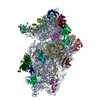 5k0yC M: map data used to model this data C: citing same article ( |
|---|---|
| Similar structure data |
-Links
 PDBj
PDBj


- Assembly
| Deposited unit | 
|
|---|---|
| 1 |
|
-Components
| #1: Protein | Mass: 66975.109 Da / Num. of mol.: 1 / Fragment: UNP Residues 170-745 Source method: isolated from a genetically manipulated source Source: (gene. exp.) Homo sapiens (human) / Gene: EIF3B, EIF3S9 / Production host: Homo sapiens (human) / References: UniProt: P55884 |
|---|---|
| #2: Protein | Mass: 4613.678 Da / Num. of mol.: 1 Source method: isolated from a genetically manipulated source Source: (gene. exp.) |
-Experimental details
-Experiment
| Experiment | Method: ELECTRON MICROSCOPY |
|---|---|
| EM experiment | Aggregation state: PARTICLE / 3D reconstruction method: single particle reconstruction |
- Sample preparation
| Component | Name: Structure of a partial yeast 48S preinitiation complex in closed conformation. Type: COMPLEX / Entity ID: all / Source: MULTIPLE SOURCES |
|---|---|
| Molecular weight | Value: 116 kDa/nm / Experimental value: NO |
| Buffer solution | pH: 6.5 |
| Specimen | Embedding applied: NO / Shadowing applied: NO / Staining applied: NO / Vitrification applied: YES |
| Vitrification | Cryogen name: ETHANE |
- Electron microscopy imaging
| Experimental equipment |  Model: Titan Krios / Image courtesy: FEI Company |
|---|---|
| Microscopy | Model: FEI TITAN KRIOS |
| Electron gun | Electron source:  FIELD EMISSION GUN / Accelerating voltage: 300 kV / Illumination mode: FLOOD BEAM FIELD EMISSION GUN / Accelerating voltage: 300 kV / Illumination mode: FLOOD BEAM |
| Electron lens | Mode: BRIGHT FIELD |
| Image recording | Electron dose: 27 e/Å2 / Film or detector model: FEI FALCON II (4k x 4k) |
- Processing
| EM software |
| |||||||||
|---|---|---|---|---|---|---|---|---|---|---|
| CTF correction | Type: NONE | |||||||||
| 3D reconstruction | Resolution: 4.9 Å / Resolution method: FSC 0.143 CUT-OFF / Num. of particles: 254957 / Symmetry type: POINT | |||||||||
| Atomic model building | Protocol: FLEXIBLE FIT / Details: MDFF | |||||||||
| Atomic model building | 3D fitting-ID: 1 / Accession code: 5A5U / Initial refinement model-ID: 1 / PDB-ID: 5A5U / Source name: PDB / Type: experimental model
|