 Movie
Movie Controller


+ Open data

- Basic information
| Entry | Database: PDB / ID: 5jws | ||||||
|---|---|---|---|---|---|---|---|
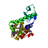
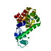
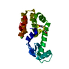
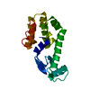
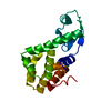
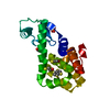
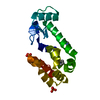
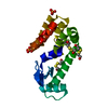
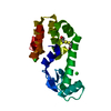
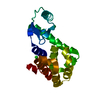
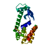
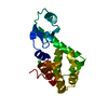
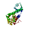
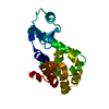
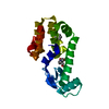
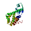
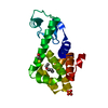
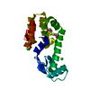
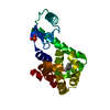
| Title | T4 Lysozyme L99A with 1-Hydro-2-ethyl-1,2-azaborine Bound | ||||||
 Components Components | Endolysin | ||||||
 Keywords Keywords | HYDROLASE / Phage lysozyme Azaborine | ||||||
| Function / homology |  Function and homology information Function and homology informationviral release from host cell by cytolysis / peptidoglycan catabolic process / cell wall macromolecule catabolic process / lysozyme / lysozyme activity / host cell cytoplasm / defense response to bacterium Similarity search - Function | ||||||
| Biological species |  Enterobacteria phage T4 sensu lato (virus) Enterobacteria phage T4 sensu lato (virus) | ||||||
| Method |  X-RAY DIFFRACTION / SYNCHROTRON / MOLECULAR REPLACEMENT / Resolution: 1.65 Å X-RAY DIFFRACTION / SYNCHROTRON / MOLECULAR REPLACEMENT / Resolution: 1.65 Å | ||||||
 Authors Authors | Lee, H. / Fischer, M. / Shoichet, B.K. / Liu, S.-Y. | ||||||
| Funding support |  United States, 1items United States, 1items
| ||||||
 Citation Citation | Journal: J.Am.Chem.Soc. / Year: 2016 Title: Hydrogen Bonding of 1,2-Azaborines in the Binding Cavity of T4 Lysozyme Mutants: Structures and Thermodynamics. Authors: Lee, H. / Fischer, M. / Shoichet, B.K. / Liu, S.Y. | ||||||
| History |
|
- Structure visualization
| Structure viewer | Molecule: MolmilJmol/JSmol |
|---|
- Downloads & links
-Download
| PDBx/mmCIF format | 5jws.cif.gz | 58.2 KB | Display | PDBx/mmCIF format |
|---|---|---|---|---|
| PDB format | pdb5jws.ent.gz | 40.1 KB | Display | PDB format |
| PDBx/mmJSON format | 5jws.json.gz | Tree view | PDBx/mmJSON format | |
| Others |  Other downloads Other downloads |
-Validation report
| Arichive directory | https://data.pdbj.org/pub/pdb/validation_reports/jw/5jwsftp://data.pdbj.org/pub/pdb/validation_reports/jw/5jws | HTTPS FTP |
|---|
-Related structure data
| Related structure data | 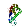 5jwtC 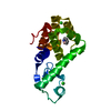 5jwuC 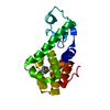 5jwvC 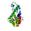 5jwwC 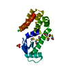 3dmvS  5jwx C: citing same article ( S: Starting model for refinement |
|---|---|
| Similar structure data |

-Links
 PDBj
PDBj


- Assembly
| Deposited unit | 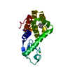
| ||||||||
|---|---|---|---|---|---|---|---|---|---|
| 1 |
| ||||||||
| Unit cell |
| ||||||||
| Components on special symmetry positions |
|
-Components
| #1: Protein | Mass: 18586.283 Da / Num. of mol.: 1 Source method: isolated from a genetically manipulated source Source: (gene. exp.) Enterobacteria phage T4 sensu lato (virus)Production host:  | ||
|---|---|---|---|
| #2: Chemical | ChemComp-6OQ /   Mass: 106.961 Da / Num. of mol.: 1 / Source method: obtained synthetically / Formula: C6H10BN Mass: 106.961 Da / Num. of mol.: 1 / Source method: obtained synthetically / Formula: C6H10BN | ||
| #3: Chemical |   Mass: 35.453 Da / Num. of mol.: 2 / Source method: obtained synthetically / Formula: Cl Mass: 35.453 Da / Num. of mol.: 2 / Source method: obtained synthetically / Formula: Cl#4: Water | ChemComp-HOH / |  Mass: 18.015 Da / Num. of mol.: 321 / Source method: isolated from a natural source / Formula: H2O Mass: 18.015 Da / Num. of mol.: 321 / Source method: isolated from a natural source / Formula: H2O |
-Experimental details
-Experiment
| Experiment | Method: X-RAY DIFFRACTION / Number of used crystals: 1 |
|---|
- Sample preparation
| Crystal | Density Matthews: 2.74 Å3/Da / Density % sol: 55.06 % |
|---|---|
| Crystal grow | Temperature: 277.15 K / Method: vapor diffusion, sitting drop / pH: 7 Details: 2.2 M sodium/potassium phosphate, pH 7.0, 50 mM 2-mercaptoethanol, 50 mM 2-hydroxyethyl disulfide |
-Data collection
| Diffraction | Mean temperature: 100 K |
|---|---|
| Diffraction source | Source: SYNCHROTRON / Site: ALS / Beamline: 8.3.1 / Wavelength: 1.11587 Å |
| Detector | Type: ADSC QUANTUM 315r / Detector: CCD / Date: Jun 3, 2015 |
| Radiation | Monochromator: KHOZU Double flat crystal / Protocol: SINGLE WAVELENGTH / Monochromatic (M) / Laue (L): M / Scattering type: x-ray |
| Radiation wavelength | Wavelength: 1.11587 Å / Relative weight: 1 |
| Reflection | Resolution: 1.65→31.87 Å / Num. obs: 23693 / % possible obs: 96 % / Redundancy: 8 % / Rmerge(I) obs: 0.118 / Net I/σ(I): 3.3 |
| Reflection shell | Resolution: 1.65→1.71 Å / Rmerge(I) obs: 0.294 / % possible all: 99.9 |
- Processing
| Software |
| ||||||||||||||||||||||||||||||||||||||||||||||||||||||||||||||||||||||
|---|---|---|---|---|---|---|---|---|---|---|---|---|---|---|---|---|---|---|---|---|---|---|---|---|---|---|---|---|---|---|---|---|---|---|---|---|---|---|---|---|---|---|---|---|---|---|---|---|---|---|---|---|---|---|---|---|---|---|---|---|---|---|---|---|---|---|---|---|---|---|---|
| Refinement | Method to determine structure: MOLECULAR REPLACEMENT Starting model: 3DMV Resolution: 1.65→31.87 Å / SU ML: 0.22 / Cross valid method: FREE R-VALUE / σ(F): 1.46 / Phase error: 24.44
| ||||||||||||||||||||||||||||||||||||||||||||||||||||||||||||||||||||||
| Solvent computation | Shrinkage radii: 0.9 Å / VDW probe radii: 1.11 Å | ||||||||||||||||||||||||||||||||||||||||||||||||||||||||||||||||||||||
| Displacement parameters | Biso max: 63.9 Å2 / Biso mean: 18.1885 Å2 / Biso min: 6.36 Å2 | ||||||||||||||||||||||||||||||||||||||||||||||||||||||||||||||||||||||
| Refinement step | Cycle: final / Resolution: 1.65→31.87 Å
| ||||||||||||||||||||||||||||||||||||||||||||||||||||||||||||||||||||||
| Refine LS restraints |
| ||||||||||||||||||||||||||||||||||||||||||||||||||||||||||||||||||||||
| LS refinement shell | Refine-ID: X-RAY DIFFRACTION / Total num. of bins used: 9
|
