 Movie
Movie Controller


+ Open data

- Basic information
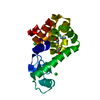
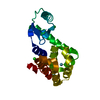
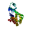
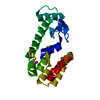
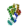
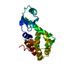
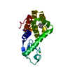
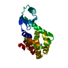
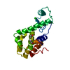
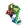
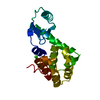
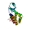
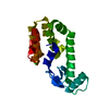
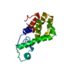
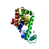
| Entry | Database: PDB / ID: 3huq | ||||||
|---|---|---|---|---|---|---|---|
| Title | Thieno[3,2-b]thiophene in complex with T4 lysozyme L99A/M102Q | ||||||
 Components Components | Lysozyme | ||||||
 Keywords Keywords | HYDROLASE / GLYCOSIDASE / BACTERIOLYTIC ENZYME / Antimicrobial | ||||||
| Function / homology |  Function and homology information Function and homology informationviral release from host cell by cytolysis / peptidoglycan catabolic process / cell wall macromolecule catabolic process / lysozyme / lysozyme activity / host cell cytoplasm / defense response to bacterium Similarity search - Function | ||||||
| Biological species |  Enterobacteria phage T4 (virus) Enterobacteria phage T4 (virus) | ||||||
| Method |  X-RAY DIFFRACTION / SYNCHROTRON / REFMAC / Resolution: 1.45 Å X-RAY DIFFRACTION / SYNCHROTRON / REFMAC / Resolution: 1.45 Å | ||||||
 Authors Authors | Boyce, S.E. / Mobley, D.L. / Rocklin, G.J. / Graves, A.P. / Dill, K.A. / Shoichet, B.K. | ||||||
 Citation Citation | Journal: J.Mol.Biol. / Year: 2009 Title: Predicting ligand binding affinity with alchemical free energy methods in a polar model binding site. Authors: Boyce, S.E. / Mobley, D.L. / Rocklin, G.J. / Graves, A.P. / Dill, K.A. / Shoichet, B.K. | ||||||
| History |
|
- Structure visualization
| Structure viewer | Molecule: MolmilJmol/JSmol |
|---|
- Downloads & links
-Download
| PDBx/mmCIF format | 3huq.cif.gz | 55.2 KB | Display | PDBx/mmCIF format |
|---|---|---|---|---|
| PDB format | pdb3huq.ent.gz | 38.4 KB | Display | PDB format |
| PDBx/mmJSON format | 3huq.json.gz | Tree view | PDBx/mmJSON format | |
| Others |  Other downloads Other downloads |
-Validation report
| Arichive directory | https://data.pdbj.org/pub/pdb/validation_reports/hu/3huqftp://data.pdbj.org/pub/pdb/validation_reports/hu/3huq | HTTPS FTP |
|---|
-Related structure data
| Related structure data | 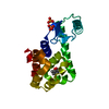 3ht6C 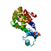 3ht7C 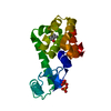 3ht8C 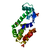 3ht9C 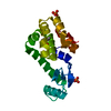 3htbC 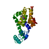 3htdC 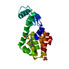 3htfC 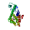 3htgC 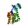 3hu8C 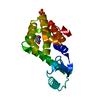 3hu9C 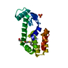 3huaC 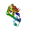 3hukC 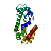 1lguS S: Starting model for refinement C: citing same article ( |
|---|---|
| Similar structure data |


-Links
 PDBj
PDBj


- Assembly
| Deposited unit | 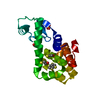
| ||||||||
|---|---|---|---|---|---|---|---|---|---|
| 1 |
| ||||||||
| Unit cell |
|
-Components
| #1: Protein | Mass: 18419.057 Da / Num. of mol.: 1 / Mutation: S38D,L99A,M102Q,N144D Source method: isolated from a genetically manipulated source Source: (gene. exp.) Enterobacteria phage T4 (virus) / Strain: Enterobacteria Phage T4 Sensu Lato / Gene: E / Plasmid: M13 / Production host:  | ||
|---|---|---|---|
| #2: Chemical | ChemComp-PO4 /   Mass: 94.971 Da / Num. of mol.: 1 / Source method: obtained synthetically / Formula: PO4 Mass: 94.971 Da / Num. of mol.: 1 / Source method: obtained synthetically / Formula: PO4 | ||
| #3: Chemical | ChemComp-J1Z / 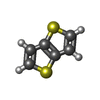  Mass: 140.226 Da / Num. of mol.: 1 / Source method: obtained synthetically / Formula: C6H4S2 Mass: 140.226 Da / Num. of mol.: 1 / Source method: obtained synthetically / Formula: C6H4S2 | ||
| #4: Chemical |   Mass: 78.133 Da / Num. of mol.: 3 / Source method: obtained synthetically / Formula: C2H6OS Mass: 78.133 Da / Num. of mol.: 3 / Source method: obtained synthetically / Formula: C2H6OS#5: Water | ChemComp-HOH / |  Mass: 18.015 Da / Num. of mol.: 278 / Source method: isolated from a natural source / Formula: H2O Mass: 18.015 Da / Num. of mol.: 278 / Source method: isolated from a natural source / Formula: H2O |
-Experimental details
-Experiment
| Experiment | Method: X-RAY DIFFRACTION / Number of used crystals: 1 |
|---|
- Sample preparation
| Crystal | Density Matthews: 2.74 Å3/Da / Density % sol: 55.14 % |
|---|---|
| Crystal grow | Temperature: 277 K / Method: vapor diffusion, hanging drop / pH: 6.5 Details: 2.2M sodium-potassium phosphate, 0.05M beta-mercaptoethanol, 0.05M 2-hydroxyethyldisulfide, pH 6.5, vapor diffusion, hanging drop, temperature 277K |
-Data collection
| Diffraction | Mean temperature: 296 K |
|---|---|
| Diffraction source | Source: SYNCHROTRON / Site: ALS  / Beamline: 8.3.1 / Wavelength: 1.11589 Å / Beamline: 8.3.1 / Wavelength: 1.11589 Å |
| Detector | Type: ADSC QUANTUM 315r / Detector: CCD / Date: Jan 27, 2008 |
| Radiation | Protocol: SINGLE WAVELENGTH / Monochromatic (M) / Laue (L): M / Scattering type: x-ray |
| Radiation wavelength | Wavelength: 1.11589 Å / Relative weight: 1 |
| Reflection | Resolution: 1.45→30 Å / Num. all: 36452 / Num. obs: 36452 / % possible obs: 99.7 % / Observed criterion σ(F): 0 / Observed criterion σ(I): 0 / Redundancy: 28.71 % / Biso Wilson estimate: 17.051 Å2 / Rmerge(I) obs: 0.048 / Net I/σ(I): 53.13 |
| Reflection shell | Resolution: 1.45→1.54 Å / Redundancy: 26.62 % / Rmerge(I) obs: 0.143 / Mean I/σ(I) obs: 24.5 / Num. measured obs: 174474 / Num. unique all: 5891 / Num. unique obs: 5891 / % possible all: 98.7 |
- Processing
| Software |
| ||||||||||||||||||||||||||||||||||||||||||||||||||||||||||||||||||||||||||||||||||||||||||
|---|---|---|---|---|---|---|---|---|---|---|---|---|---|---|---|---|---|---|---|---|---|---|---|---|---|---|---|---|---|---|---|---|---|---|---|---|---|---|---|---|---|---|---|---|---|---|---|---|---|---|---|---|---|---|---|---|---|---|---|---|---|---|---|---|---|---|---|---|---|---|---|---|---|---|---|---|---|---|---|---|---|---|---|---|---|---|---|---|---|---|---|
| Refinement | Method to determine structure: REFMAC Starting model: PDB ENTRY 1LGU Resolution: 1.45→27.46 Å / Cor.coef. Fo:Fc: 0.958 / Cor.coef. Fo:Fc free: 0.946 / Occupancy max: 1 / Occupancy min: 0.2 / SU B: 0.894 / SU ML: 0.036 / Cross valid method: THROUGHOUT / σ(F): 0 / ESU R: 0.061 / ESU R Free: 0.061 / Stereochemistry target values: MAXIMUM LIKELIHOOD / Details: HYDROGENS HAVE BEEN ADDED IN THE RIDING POSITIONS
| ||||||||||||||||||||||||||||||||||||||||||||||||||||||||||||||||||||||||||||||||||||||||||
| Solvent computation | Ion probe radii: 0.8 Å / Shrinkage radii: 0.8 Å / VDW probe radii: 1.2 Å / Solvent model: MASK | ||||||||||||||||||||||||||||||||||||||||||||||||||||||||||||||||||||||||||||||||||||||||||
| Displacement parameters | Biso max: 48.13 Å2 / Biso mean: 11.731 Å2 / Biso min: 2 Å2
| ||||||||||||||||||||||||||||||||||||||||||||||||||||||||||||||||||||||||||||||||||||||||||
| Refinement step | Cycle: LAST / Resolution: 1.45→27.46 Å
| ||||||||||||||||||||||||||||||||||||||||||||||||||||||||||||||||||||||||||||||||||||||||||
| Refine LS restraints |
| ||||||||||||||||||||||||||||||||||||||||||||||||||||||||||||||||||||||||||||||||||||||||||
| LS refinement shell | Resolution: 1.45→1.489 Å / Total num. of bins used: 20
|
