 Movie
Movie Controller


+ Open data

- Basic information
| Entry | Database: PDB / ID: 2ray | ||||||
|---|---|---|---|---|---|---|---|
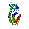
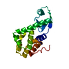
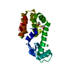
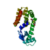
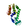
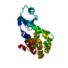
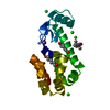
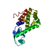
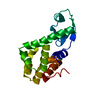
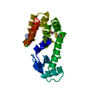
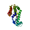
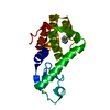
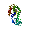
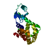
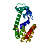
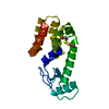
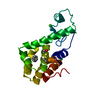
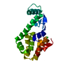
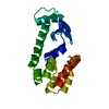
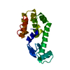
| Title | beta-chlorophenetole in complex with T4 lysozyme L99A | ||||||
 Components Components | Lysozyme | ||||||
 Keywords Keywords | HYDROLASE / protein cavities | ||||||
| Function / homology |  Function and homology information Function and homology informationviral release from host cell by cytolysis / peptidoglycan catabolic process / cell wall macromolecule catabolic process / lysozyme / lysozyme activity / host cell cytoplasm / defense response to bacterium Similarity search - Function | ||||||
| Biological species |  Enterobacteria phage T4 (virus) Enterobacteria phage T4 (virus) | ||||||
| Method |  X-RAY DIFFRACTION / Resolution: 1.798 Å X-RAY DIFFRACTION / Resolution: 1.798 Å | ||||||
 Authors Authors | Graves, A.P. / Boyce, S.E. / Shoichet, B.K. | ||||||
 Citation Citation | Journal: J.Mol.Biol. / Year: 2008 Title: Rescoring docking hit lists for model cavity sites: predictions and experimental testing. Authors: Graves, A.P. / Shivakumar, D.M. / Boyce, S.E. / Jacobson, M.P. / Case, D.A. / Shoichet, B.K. | ||||||
| History |
|
- Structure visualization
| Structure viewer | Molecule: MolmilJmol/JSmol |
|---|
- Downloads & links
-Download
| PDBx/mmCIF format | 2ray.cif.gz | 51.2 KB | Display | PDBx/mmCIF format |
|---|---|---|---|---|
| PDB format | pdb2ray.ent.gz | 35.4 KB | Display | PDB format |
| PDBx/mmJSON format | 2ray.json.gz | Tree view | PDBx/mmJSON format | |
| Others |  Other downloads Other downloads |
-Validation report
| Arichive directory | https://data.pdbj.org/pub/pdb/validation_reports/ra/2rayftp://data.pdbj.org/pub/pdb/validation_reports/ra/2ray | HTTPS FTP |
|---|
-Related structure data
| Related structure data | 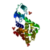 2razC 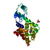 2rb0C 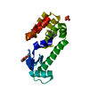 2rb1C 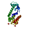 2rb2C 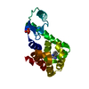 2rbnC 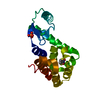 2rboC 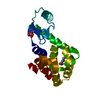 2rbpC 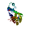 2rbqC  2rbrC  2rbsC  2rbtC  2rbuC  2rbvC  2rbwC 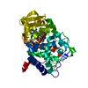 2rbxC 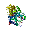 2rbyC  2rbzC 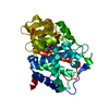 2rc0C 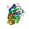 2rc1C  2rc2C 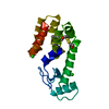 184lS C: citing same article ( S: Starting model for refinement |
|---|---|
| Similar structure data |


-Links
 PDBj
PDBj


- Assembly
| Deposited unit | 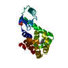
| ||||||||
|---|---|---|---|---|---|---|---|---|---|
| 1 |
| ||||||||
| Unit cell |
|
-Components
| #1: Protein | Mass: 18359.023 Da / Num. of mol.: 1 / Mutation: L99A Source method: isolated from a genetically manipulated source Source: (gene. exp.) Enterobacteria phage T4 (virus) / Genus: T4-like viruses / Species: Enterobacteria phage T4 sensu lato / Gene: E / Plasmid: M13 / Production host:  |
|---|---|
| #2: Chemical | ChemComp-PO4 /   Mass: 94.971 Da / Num. of mol.: 1 / Source method: obtained synthetically / Formula: PO4 Mass: 94.971 Da / Num. of mol.: 1 / Source method: obtained synthetically / Formula: PO4 |
| #3: Chemical | ChemComp-258 / (  Mass: 156.609 Da / Num. of mol.: 1 / Source method: obtained synthetically / Formula: C8H9ClO Mass: 156.609 Da / Num. of mol.: 1 / Source method: obtained synthetically / Formula: C8H9ClO |
| #4: Water | ChemComp-HOH /  Mass: 18.015 Da / Num. of mol.: 212 / Source method: isolated from a natural source / Formula: H2O Mass: 18.015 Da / Num. of mol.: 212 / Source method: isolated from a natural source / Formula: H2O |
-Experimental details
-Experiment
| Experiment | Method: X-RAY DIFFRACTION / Number of used crystals: 1 |
|---|
- Sample preparation
| Crystal | Density Matthews: 2.76 Å3/Da / Density % sol: 55.41 % |
|---|---|
| Crystal grow | Temperature: 277 K / Method: hanging drop / pH: 7.1 Details: 2.2M sodium-potassium phosphate, 0.05M beta-mercaptoethanol, 0.05M 2-hydroxyethyldisulfide, pH 7.1, hanging drop, temperature 277K |
-Data collection
| Diffraction | Mean temperature: 110 K |
|---|---|
| Diffraction source | Source: ROTATING ANODE / Type: RIGAKU RU200 / Wavelength: 1.5418 Å |
| Detector | Type: RIGAKU RAXIS IV / Detector: IMAGE PLATE / Date: Nov 26, 2006 |
| Radiation | Protocol: SINGLE WAVELENGTH / Monochromatic (M) / Laue (L): M / Scattering type: x-ray |
| Radiation wavelength | Wavelength: 1.5418 Å / Relative weight: 1 |
| Reflection | Resolution: 1.798→52.34 Å / Num. all: 19492 / Num. obs: 19434 / % possible obs: 99.7 % / Observed criterion σ(F): 0 / Observed criterion σ(I): 0 |
| Reflection shell | Resolution: 1.798→1.84 Å / % possible all: 98.9 |
- Processing
| Software |
| ||||||||||||||||||||||||||||||||||||||||||||||||||||||||||||||||||||||||||||||||||||||||||
|---|---|---|---|---|---|---|---|---|---|---|---|---|---|---|---|---|---|---|---|---|---|---|---|---|---|---|---|---|---|---|---|---|---|---|---|---|---|---|---|---|---|---|---|---|---|---|---|---|---|---|---|---|---|---|---|---|---|---|---|---|---|---|---|---|---|---|---|---|---|---|---|---|---|---|---|---|---|---|---|---|---|---|---|---|---|---|---|---|---|---|---|
| Refinement | Starting model: 184L Resolution: 1.798→52.34 Å / Cor.coef. Fo:Fc: 0.954 / Cor.coef. Fo:Fc free: 0.942 / SU B: 2.374 / SU ML: 0.075 / Cross valid method: THROUGHOUT / σ(F): 0 / ESU R: 0.119 / ESU R Free: 0.111 / Stereochemistry target values: MAXIMUM LIKELIHOOD / Details: HYDROGENS HAVE BEEN ADDED IN THE RIDING POSITIONS
| ||||||||||||||||||||||||||||||||||||||||||||||||||||||||||||||||||||||||||||||||||||||||||
| Solvent computation | Ion probe radii: 0.8 Å / Shrinkage radii: 0.8 Å / VDW probe radii: 1.2 Å / Solvent model: MASK | ||||||||||||||||||||||||||||||||||||||||||||||||||||||||||||||||||||||||||||||||||||||||||
| Displacement parameters | Biso mean: 20.548 Å2
| ||||||||||||||||||||||||||||||||||||||||||||||||||||||||||||||||||||||||||||||||||||||||||
| Refinement step | Cycle: LAST / Resolution: 1.798→52.34 Å
| ||||||||||||||||||||||||||||||||||||||||||||||||||||||||||||||||||||||||||||||||||||||||||
| Refine LS restraints |
| ||||||||||||||||||||||||||||||||||||||||||||||||||||||||||||||||||||||||||||||||||||||||||
| LS refinement shell | Resolution: 1.798→1.844 Å / Total num. of bins used: 20
|
