 Movie
Movie Controller


+ Open data

- Basic information
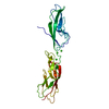
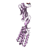
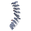
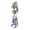
| Entry | Database: PDB / ID: 5jvm | |||||||||
|---|---|---|---|---|---|---|---|---|---|---|
| Title | The neck-linker and alpha 7 helix of Mus musculus KIF3C | |||||||||
 Components Components | Chimera protein of Kinesin-like protein KIF3C and Microtubule-associated protein RP/EB family member 1 | |||||||||
 Keywords Keywords | MOTOR PROTEIN / kinesin / coiled-coil | |||||||||
| Function / homology |  Function and homology information Function and homology informationprotein localization to astral microtubule / protein localization to mitotic spindle / Intraflagellar transport / cortical microtubule cytoskeleton / neuronal ribonucleoprotein granule / mitotic spindle astral microtubule end / Kinesins / protein localization to microtubule / COPI-dependent Golgi-to-ER retrograde traffic / cell projection membrane ...protein localization to astral microtubule / protein localization to mitotic spindle / Intraflagellar transport / cortical microtubule cytoskeleton / neuronal ribonucleoprotein granule / mitotic spindle astral microtubule end / Kinesins / protein localization to microtubule / COPI-dependent Golgi-to-ER retrograde traffic / cell projection membrane / microtubule plus-end / mitotic spindle microtubule / attachment of mitotic spindle microtubules to kinetochore / MHC class II antigen presentation / microtubule plus-end binding / microtubule bundle formation / non-motile cilium assembly / protein localization to centrosome / microtubule motor activity / microtubule-based movement / mitotic spindle pole / spindle midzone / establishment of mitotic spindle orientation / negative regulation of microtubule polymerization / microtubule polymerization / microtubule organizing center / regulation of microtubule polymerization or depolymerization / positive regulation of microtubule polymerization / cytoplasmic microtubule / spindle assembly / Loss of Nlp from mitotic centrosomes / Loss of proteins required for interphase microtubule organization from the centrosome / Amplification of signal from unattached kinetochores via a MAD2 inhibitory signal / Recruitment of mitotic centrosome proteins and complexes / Recruitment of NuMA to mitotic centrosomes / Anchoring of the basal body to the plasma membrane / Mitotic Prometaphase / EML4 and NUDC in mitotic spindle formation / AURKA Activation by TPX2 / Resolution of Sister Chromatid Cohesion / protein serine/threonine kinase binding / RHO GTPases Activate Formins / intracellular protein localization / The role of GTSE1 in G2/M progression after G2 checkpoint / Separation of Sister Chromatids / Regulation of PLK1 Activity at G2/M Transition / cell migration / microtubule cytoskeleton / microtubule binding / microtubule / ciliary basal body / cadherin binding / cell division / focal adhesion / neuronal cell body / centrosome / Golgi apparatus / RNA binding / ATP binding / identical protein binding / cytosol Similarity search - Function | |||||||||
| Biological species |   Homo sapiens (human) Homo sapiens (human) | |||||||||
| Method |  X-RAY DIFFRACTION / SYNCHROTRON / MOLECULAR REPLACEMENT / Resolution: 1.567 Å X-RAY DIFFRACTION / SYNCHROTRON / MOLECULAR REPLACEMENT / Resolution: 1.567 Å | |||||||||
 Authors Authors | Phillips, R.K. / Rayment, I. | |||||||||
| Funding support |  United States, 2items United States, 2items
| |||||||||
 Citation Citation | Journal: J.Biol.Chem. / Year: 2016 Title: Family-specific Kinesin Structures Reveal Neck-linker Length Based on Initiation of the Coiled-coil. Authors: Phillips, R.K. / Peter, L.G. / Gilbert, S.P. / Rayment, I. | |||||||||
| History |
|
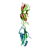
- Structure visualization
| Structure viewer | Molecule: MolmilJmol/JSmol |
|---|
- Downloads & links
-Download
| PDBx/mmCIF format | 5jvm.cif.gz | 88.5 KB | Display | PDBx/mmCIF format |
|---|---|---|---|---|
| PDB format | pdb5jvm.ent.gz | 67.4 KB | Display | PDB format |
| PDBx/mmJSON format | 5jvm.json.gz | Tree view | PDBx/mmJSON format | |
| Others |  Other downloads Other downloads |
-Validation report
| Arichive directory | https://data.pdbj.org/pub/pdb/validation_reports/jv/5jvmftp://data.pdbj.org/pub/pdb/validation_reports/jv/5jvm | HTTPS FTP |
|---|
-Related structure data
| Related structure data | 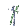 5jv3C 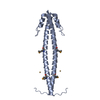 5jvpC 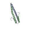 5jvrC 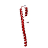 5jvsC 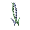 5jvuC 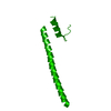 5jx1C 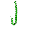 1yibS C: citing same article ( S: Starting model for refinement |
|---|---|
| Similar structure data |


-Links
 PDBj
PDBj


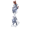
- Assembly
| Deposited unit | 
| ||||||||
|---|---|---|---|---|---|---|---|---|---|
| 1 |
| ||||||||
| Unit cell |
|
-Components
| #1: Protein | Mass: 9638.826 Da / Num. of mol.: 2 Fragment: UNP O35066 residues 374-402,UNP Q15691 residues 207-257 Source method: isolated from a genetically manipulated source Source: (gene. exp.) Homo sapiens (human)Gene: Kif3c, MAPRE1 / Production host:  #2: Chemical | ChemComp-MG / |   Mass: 24.305 Da / Num. of mol.: 1 / Source method: obtained synthetically / Formula: Mg Mass: 24.305 Da / Num. of mol.: 1 / Source method: obtained synthetically / Formula: Mg#3: Water | ChemComp-HOH / |  Mass: 18.015 Da / Num. of mol.: 276 / Source method: isolated from a natural source / Formula: H2O Mass: 18.015 Da / Num. of mol.: 276 / Source method: isolated from a natural source / Formula: H2O |
|---|
-Experimental details
-Experiment
| Experiment | Method: X-RAY DIFFRACTION / Number of used crystals: 1 |
|---|
- Sample preparation
| Crystal | Density Matthews: 2.99 Å3/Da / Density % sol: 58.88 % |
|---|---|
| Crystal grow | Temperature: 298 K / Method: vapor diffusion, hanging drop / pH: 9 Details: 30% (w/v) pentaerythritol ethoxylate (PEE) 797, 1.5% (w/v) ethylene glycol monoethylether, 400 mM MgCl2, 100 mM bis-tris propane pH 9.0 |
-Data collection
| Diffraction | Mean temperature: 100 K |
|---|---|
| Diffraction source | Source: SYNCHROTRON / Site: APS / Beamline: 19-ID / Wavelength: 0.97924 Å |
| Detector | Type: ADSC QUANTUM 315r / Detector: CCD / Date: Dec 13, 2014 |
| Radiation | Protocol: SINGLE WAVELENGTH / Monochromatic (M) / Laue (L): M / Scattering type: x-ray |
| Radiation wavelength | Wavelength: 0.97924 Å / Relative weight: 1 |
| Reflection | Resolution: 1.5667→37.004 Å / Num. obs: 44738 / % possible obs: 98 % / Redundancy: 7.1 % / Rmerge(I) obs: 0.073 / Net I/σ(I): 21.7 |
| Reflection shell | Resolution: 1.5667→1.62 Å / Redundancy: 3 % / Rmerge(I) obs: 0.595 / Mean I/σ(I) obs: 1.6 / Num. measured obs: 2513 |
- Processing
| Software |
| ||||||||||||||||||||||||||||||||||||||||||||||||||||||||||||||||||||||||||||||||||||||||||||||||||||||||||||||||||||||||||||||||||||||||||||||||||||||||||||||||||||||||||||||||||||||||||||||||||||||||
|---|---|---|---|---|---|---|---|---|---|---|---|---|---|---|---|---|---|---|---|---|---|---|---|---|---|---|---|---|---|---|---|---|---|---|---|---|---|---|---|---|---|---|---|---|---|---|---|---|---|---|---|---|---|---|---|---|---|---|---|---|---|---|---|---|---|---|---|---|---|---|---|---|---|---|---|---|---|---|---|---|---|---|---|---|---|---|---|---|---|---|---|---|---|---|---|---|---|---|---|---|---|---|---|---|---|---|---|---|---|---|---|---|---|---|---|---|---|---|---|---|---|---|---|---|---|---|---|---|---|---|---|---|---|---|---|---|---|---|---|---|---|---|---|---|---|---|---|---|---|---|---|---|---|---|---|---|---|---|---|---|---|---|---|---|---|---|---|---|---|---|---|---|---|---|---|---|---|---|---|---|---|---|---|---|---|---|---|---|---|---|---|---|---|---|---|---|---|---|---|---|---|
| Refinement | Method to determine structure: MOLECULAR REPLACEMENT Starting model: 1YIB Resolution: 1.567→37.004 Å / SU ML: 0.15 / Cross valid method: FREE R-VALUE / σ(F): 1.35 / Phase error: 22.42 / Stereochemistry target values: ML
| ||||||||||||||||||||||||||||||||||||||||||||||||||||||||||||||||||||||||||||||||||||||||||||||||||||||||||||||||||||||||||||||||||||||||||||||||||||||||||||||||||||||||||||||||||||||||||||||||||||||||
| Solvent computation | Shrinkage radii: 0.9 Å / VDW probe radii: 1.11 Å / Solvent model: FLAT BULK SOLVENT MODEL | ||||||||||||||||||||||||||||||||||||||||||||||||||||||||||||||||||||||||||||||||||||||||||||||||||||||||||||||||||||||||||||||||||||||||||||||||||||||||||||||||||||||||||||||||||||||||||||||||||||||||
| Refinement step | Cycle: LAST / Resolution: 1.567→37.004 Å
| ||||||||||||||||||||||||||||||||||||||||||||||||||||||||||||||||||||||||||||||||||||||||||||||||||||||||||||||||||||||||||||||||||||||||||||||||||||||||||||||||||||||||||||||||||||||||||||||||||||||||
| Refine LS restraints |
| ||||||||||||||||||||||||||||||||||||||||||||||||||||||||||||||||||||||||||||||||||||||||||||||||||||||||||||||||||||||||||||||||||||||||||||||||||||||||||||||||||||||||||||||||||||||||||||||||||||||||
| LS refinement shell |
| ||||||||||||||||||||||||||||||||||||||||||||||||||||||||||||||||||||||||||||||||||||||||||||||||||||||||||||||||||||||||||||||||||||||||||||||||||||||||||||||||||||||||||||||||||||||||||||||||||||||||
| Refinement TLS params. | Method: refined / Refine-ID: X-RAY DIFFRACTION
| ||||||||||||||||||||||||||||||||||||||||||||||||||||||||||||||||||||||||||||||||||||||||||||||||||||||||||||||||||||||||||||||||||||||||||||||||||||||||||||||||||||||||||||||||||||||||||||||||||||||||
| Refinement TLS group |
|
